100 years of migmatites - in Sederholm's footsteps: On migmatites and associated Precambrian rocks of SW Finland"
33rd IGC
Sederholm Symposium Field Trip August 2008
led by Olav Eklund, Carl
Ehlers and Markku Väisänen
Roberto Weinberg, Monash University, Australia
Contents
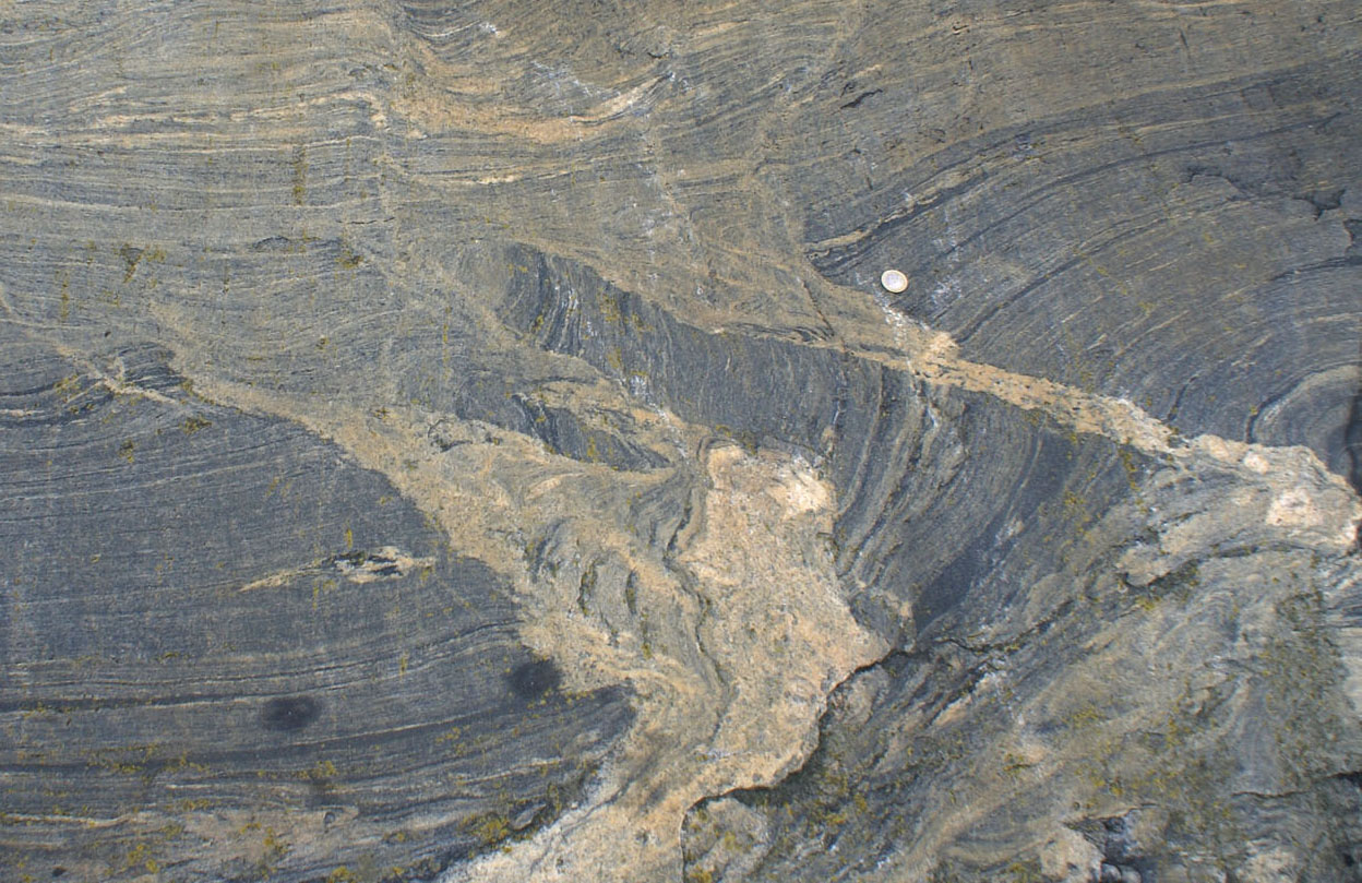
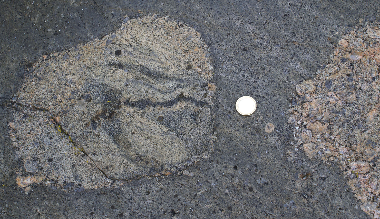
a) Cordierite-Garnet Migmatite, Masku Riviera, N of Turku (Stop 29 guidebook, N6724734, E3233489)
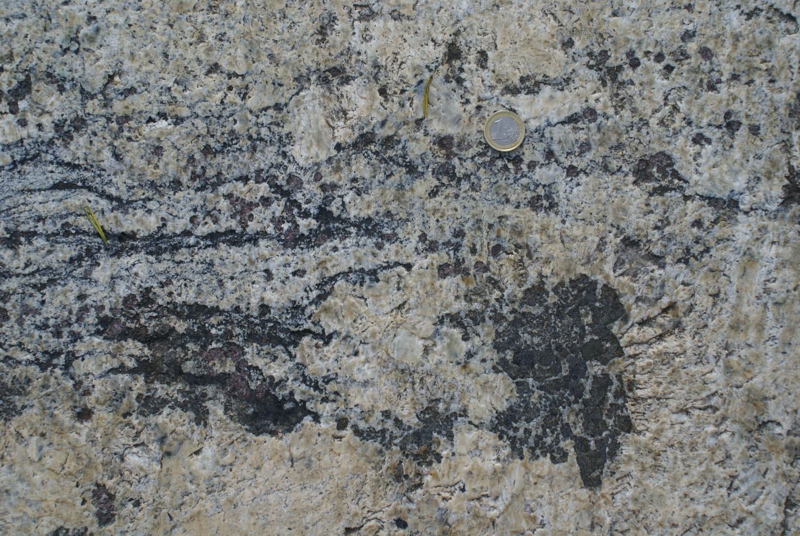
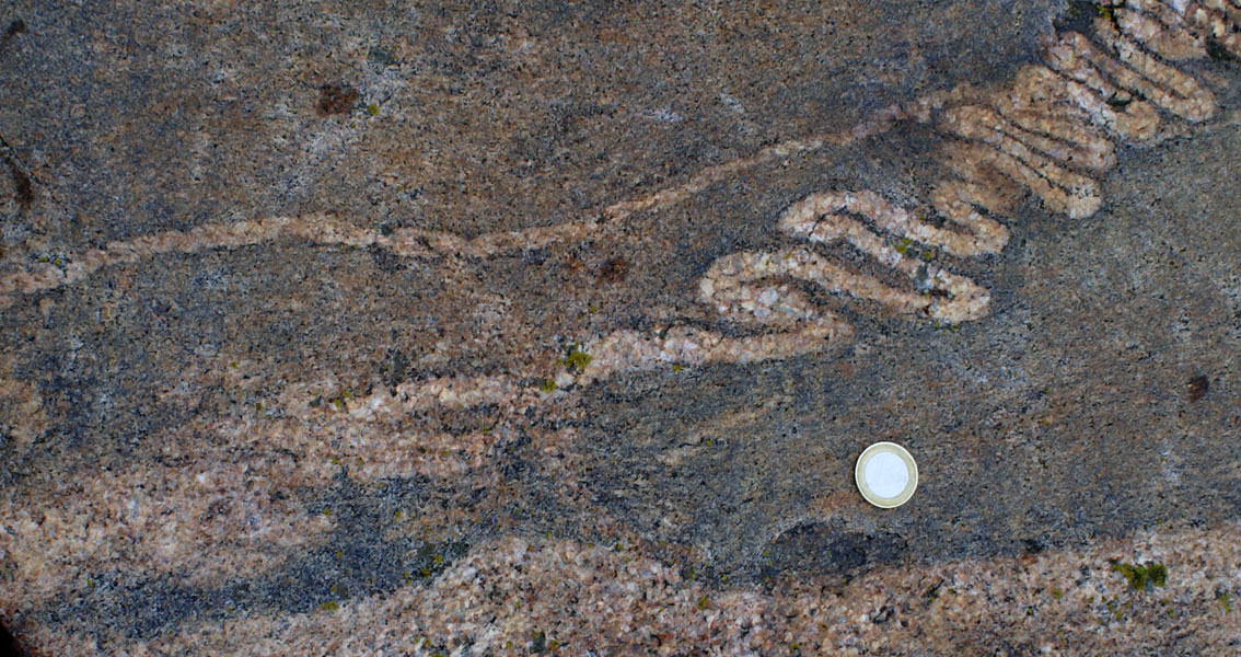
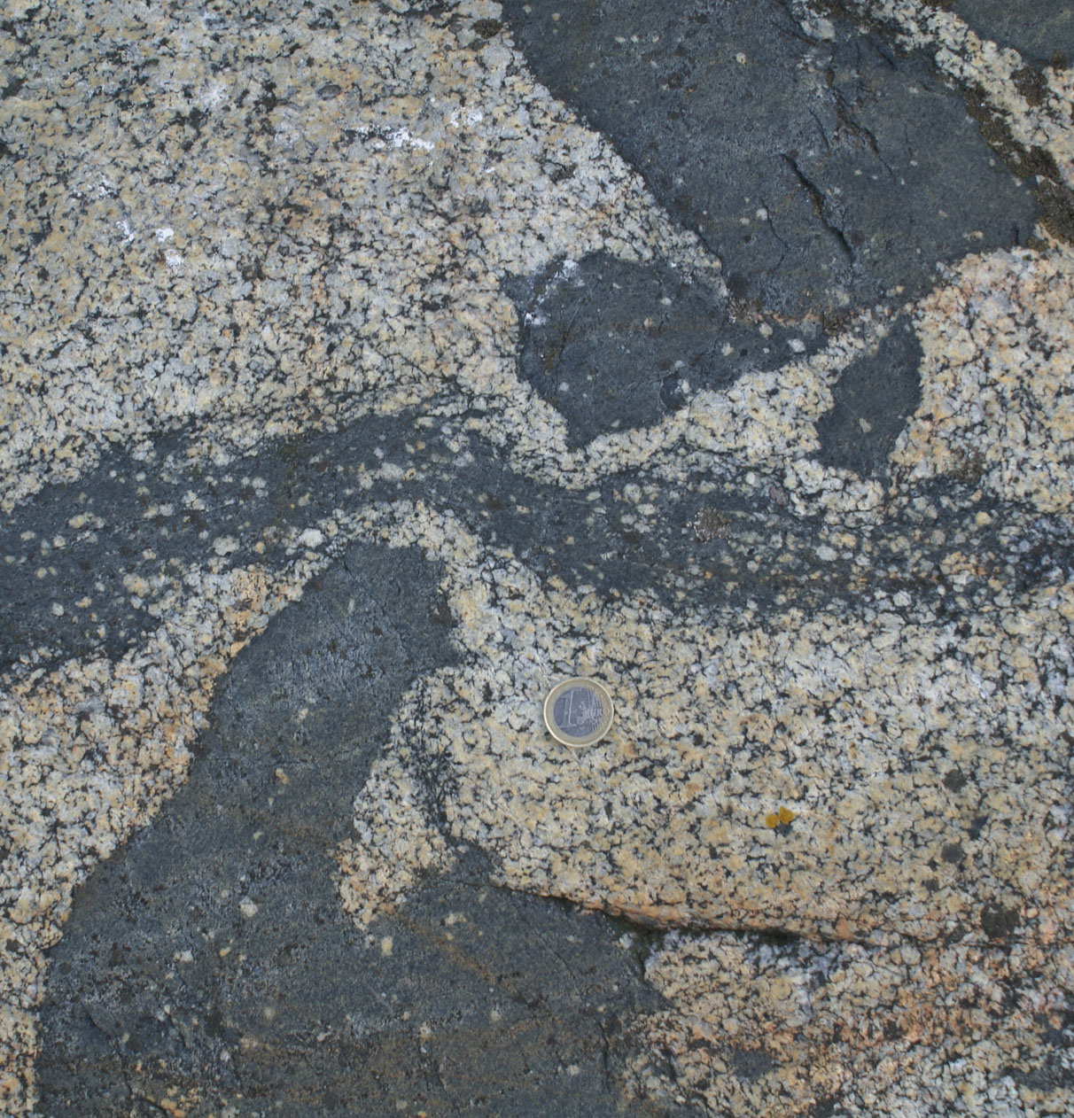
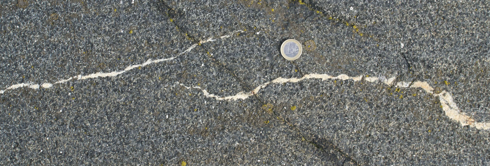
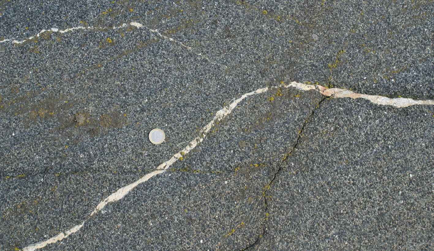
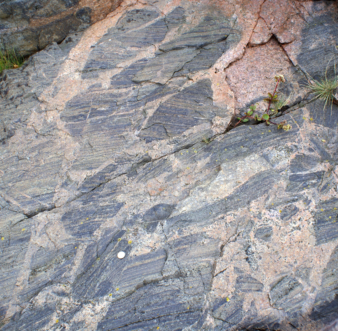
step 1. melting during folding (Fig. 1a), little melt escape through spaced axial planar leucocratic dykes (Fig. 1b)
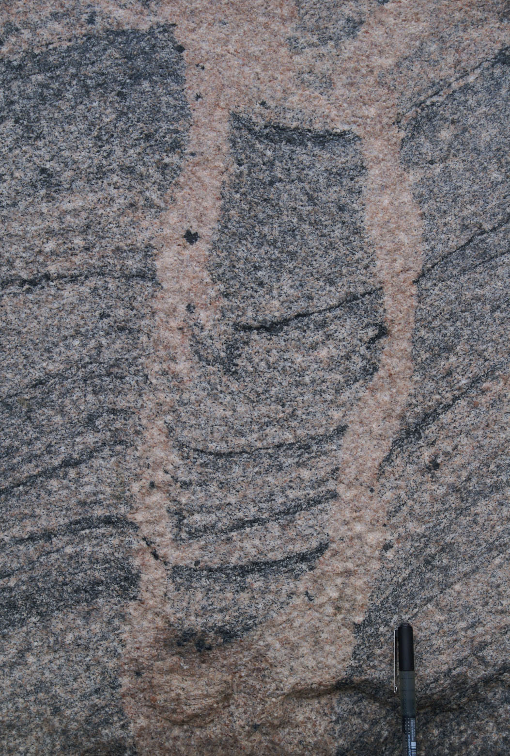
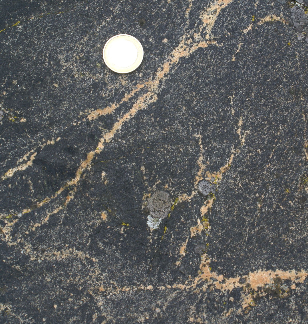
step 2. Cooling during continued folding, spaced shear bands filled-up with melt (Fig. 1c), link with (Fig. 1b) and locally displace early formed axial planar dykes

|
 |
 |
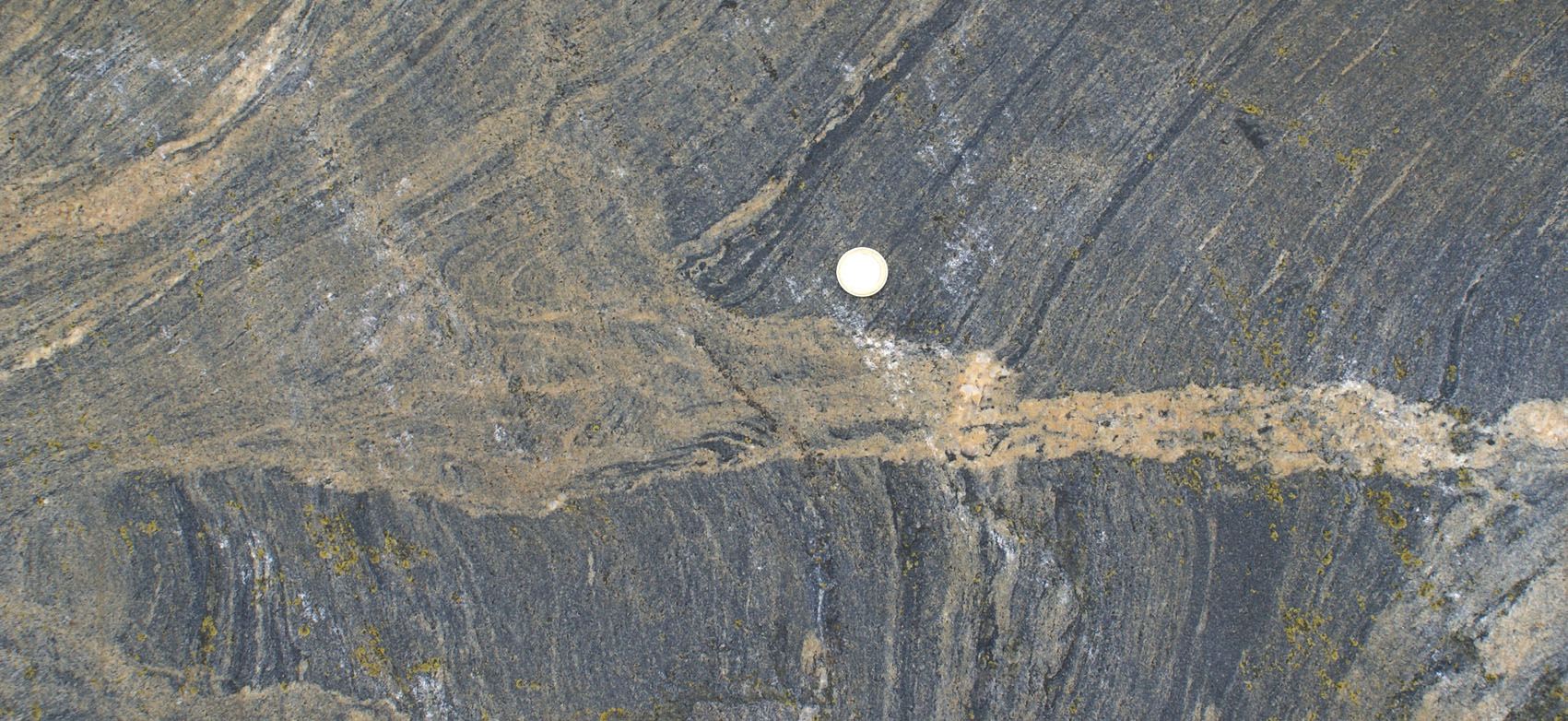
| Figure 1a) Folded migmatite. Leucosomes folded with melanosomes with no evidence at this scale for melt escape. | Figure b) Thick axial planar dykes linked directly with leucosomes, such as those folded in a). This particular example also links up with diagonal leucosomes in shear bands (above and below lens cap). | Figure c) Cordierite-garnet leucocratic dyke on shear plane including some melanocratic bands. Leucocratic material in dyke links with leucosomes in surroundings suggesting contemporaneity. |
 |

|
 |
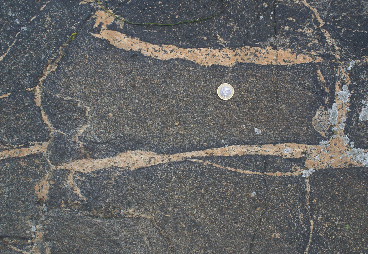
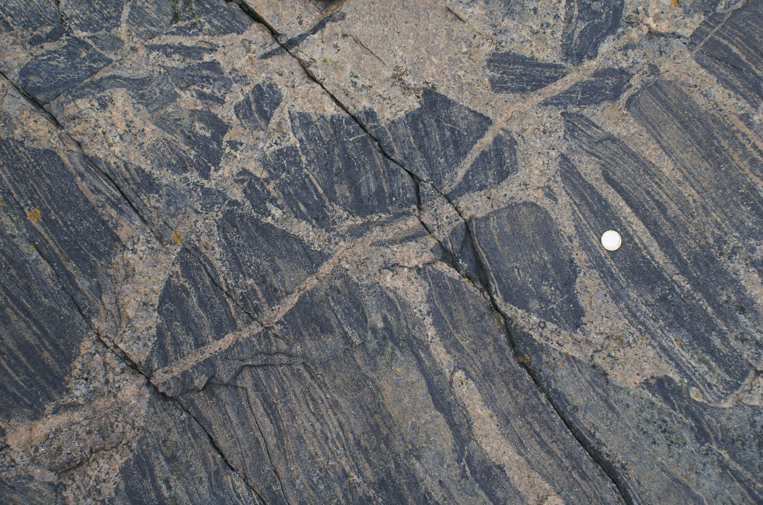
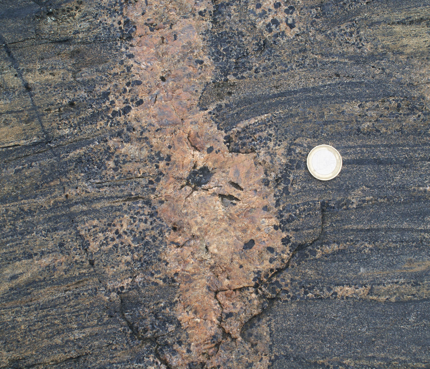
| Figure 2a) Shear plane parallel dyke carrying numerous cordierite, intrude and displace a more leucocratic axial planar dyke, indicating relative timing (click on figure to see larger version). Blue line marks the boundary. | Figure 2b) Aggregates of cordierite (dark grey grains in the lower part of the photo) and garnet in axial planar dyke with melanocratic rim. | Figure c) Same, coarse euhedral cordierite (centre) and garnet in same dyke as in b) |

|
|
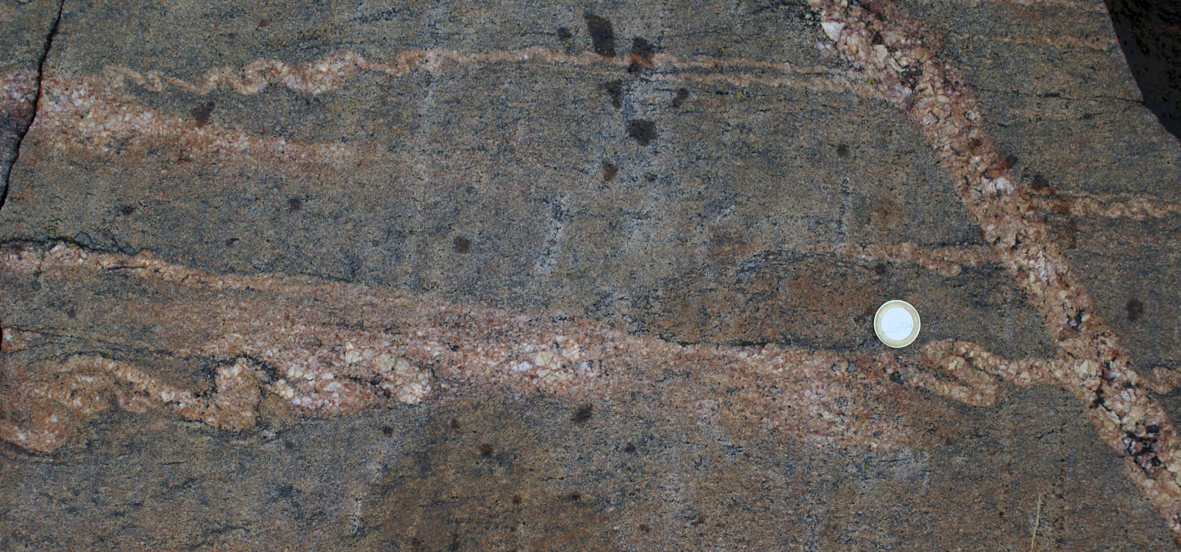
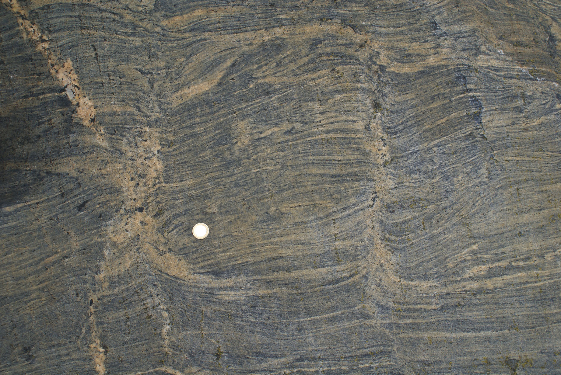
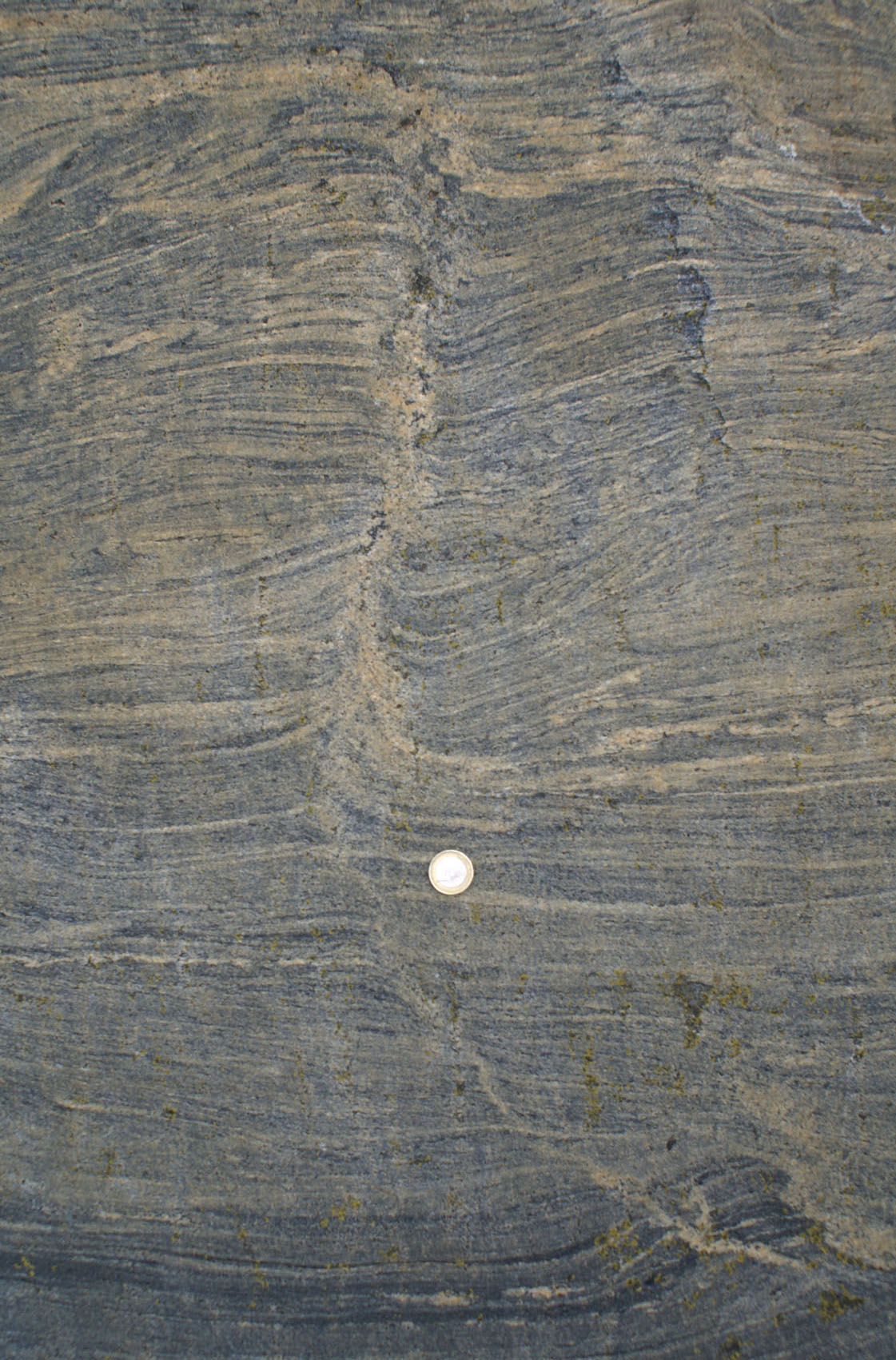
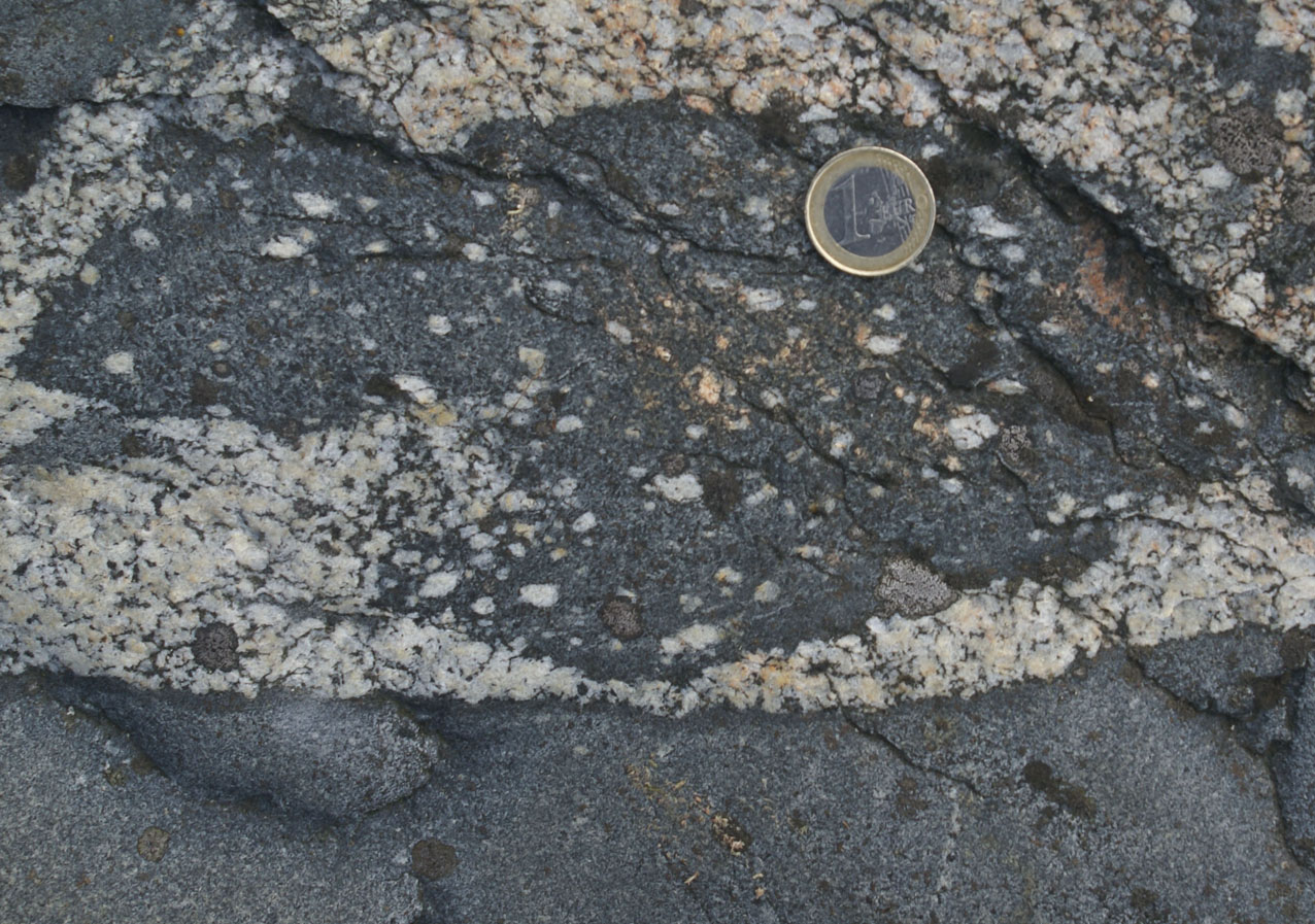
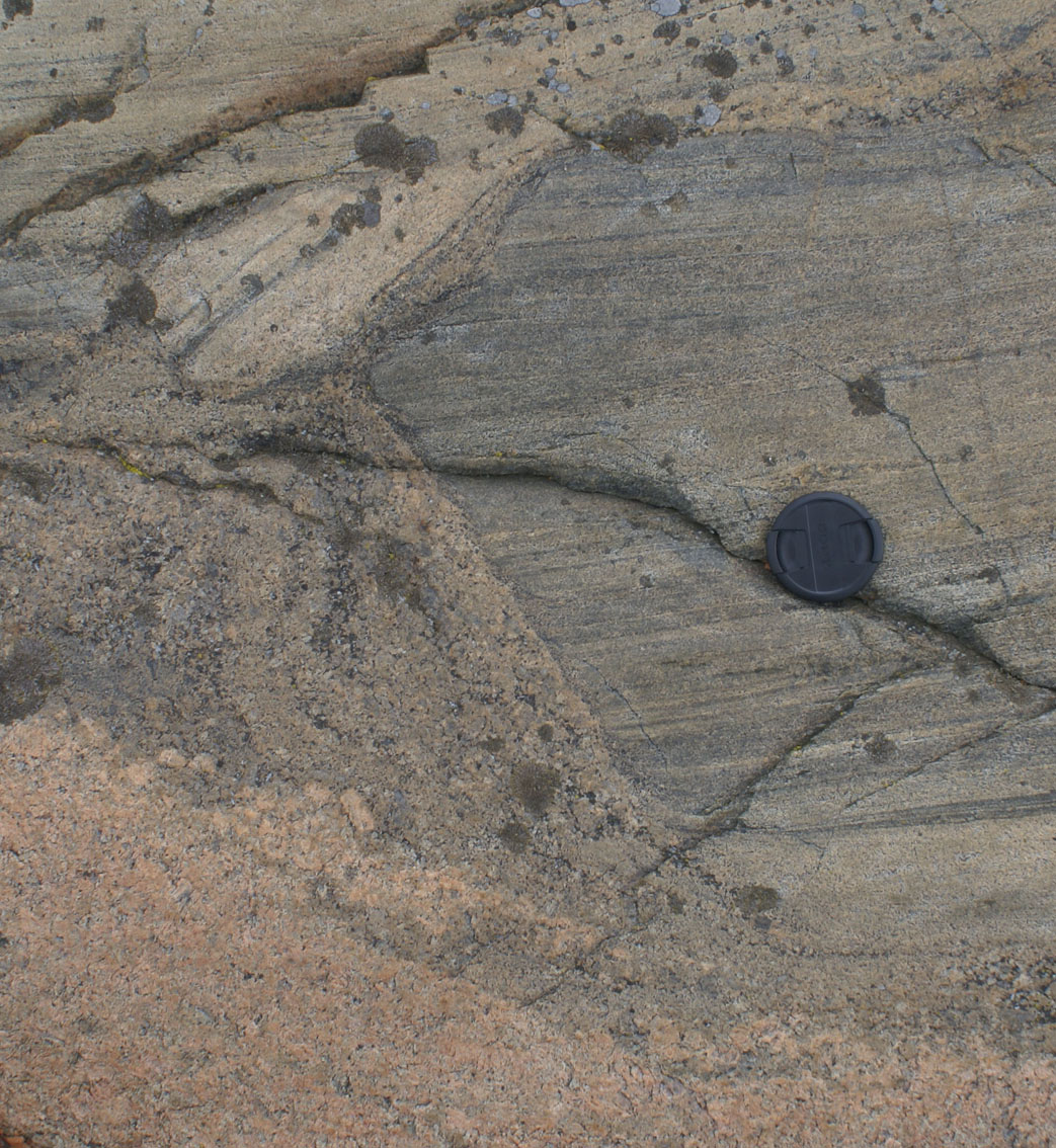
| Figure 3a) Shear plane dyke linked continuously with leucosomes in main rock fabric. Relatively little magma movement across the foliation is inferred because of preservation of delicate melanosome tips and a faint continuity of mafic minerals across the dyke diagonally from lower right to upper left. | Figure 3b) From Senate Square, Helsinki. Continuity of melanosome across axial planar dykes (with diffuse boundaries and linked with leucosomes in migmatitic gneiss) indicative of little flow along the exposed length of the dyke. |
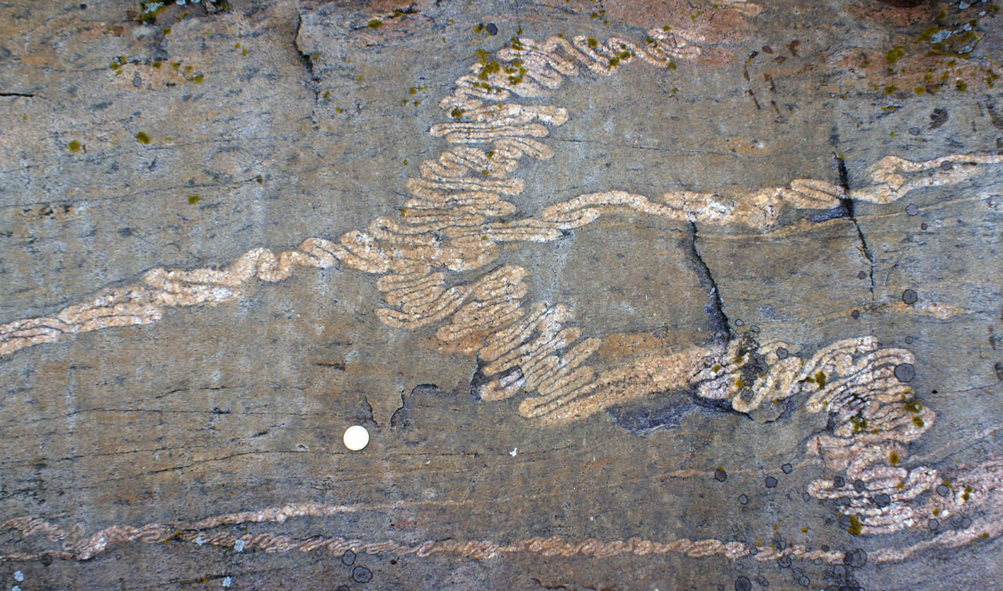
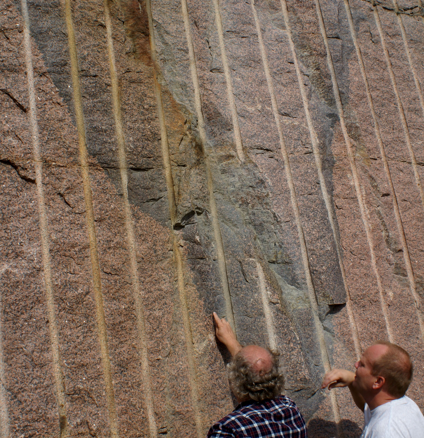
b) Ptygmatic folds and their disruption, Brändöharum (stop 3 in guidebook; N6657490, E3345404) in Sederholm 1926, Bull. Comission Geol. Finlande 77

|
|
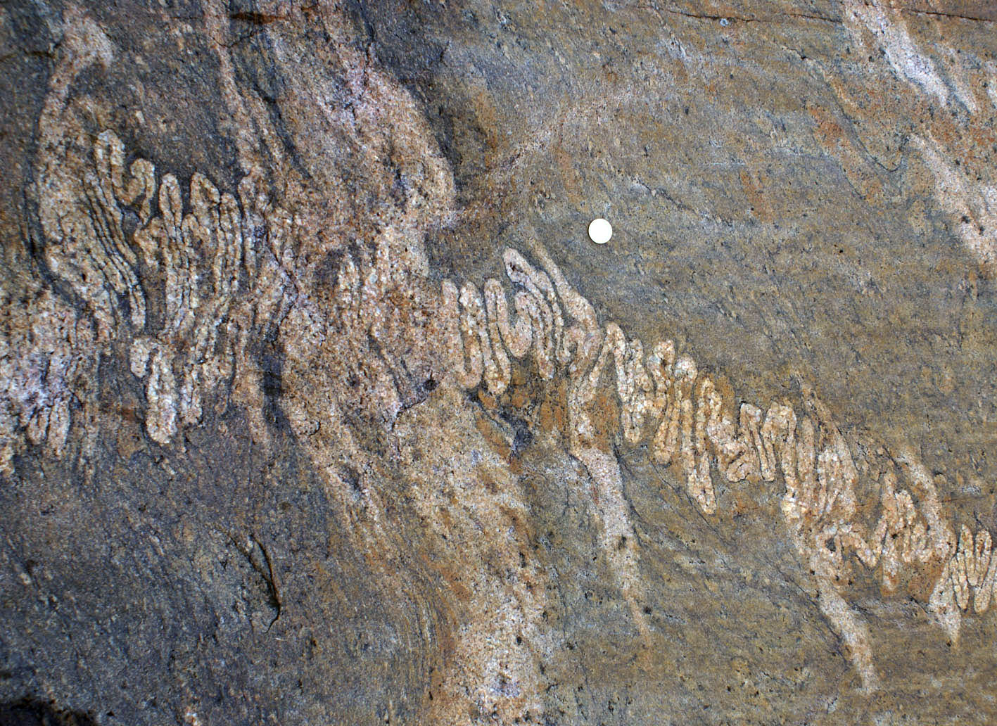
| Figure 4a, b) Several connected granitic veinlets forming ptygmatic fold trains in different orientations, looking downplunge on a horizontal plane, indicating general constriction during high-grade metamorphic conditions. |
 |
 |
|
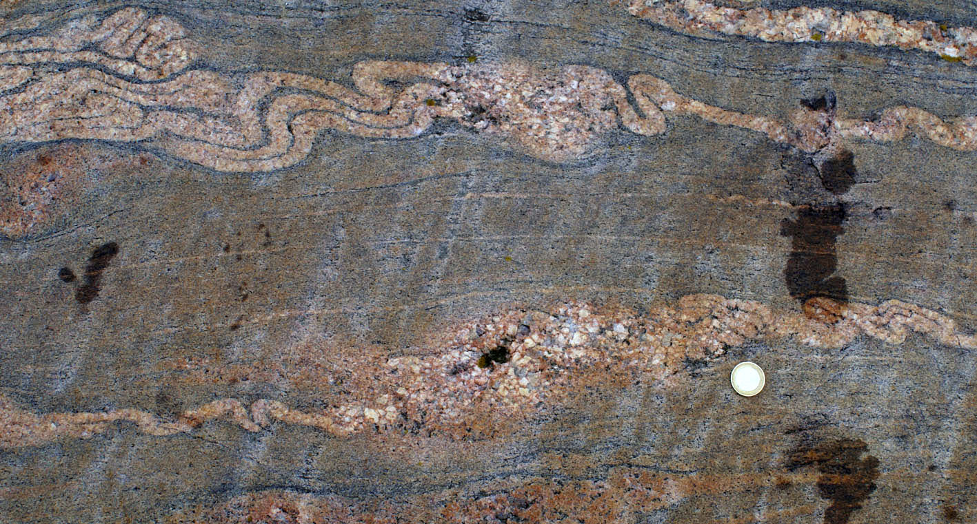
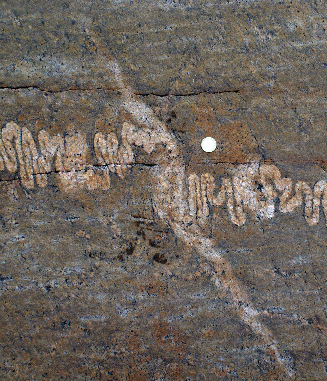
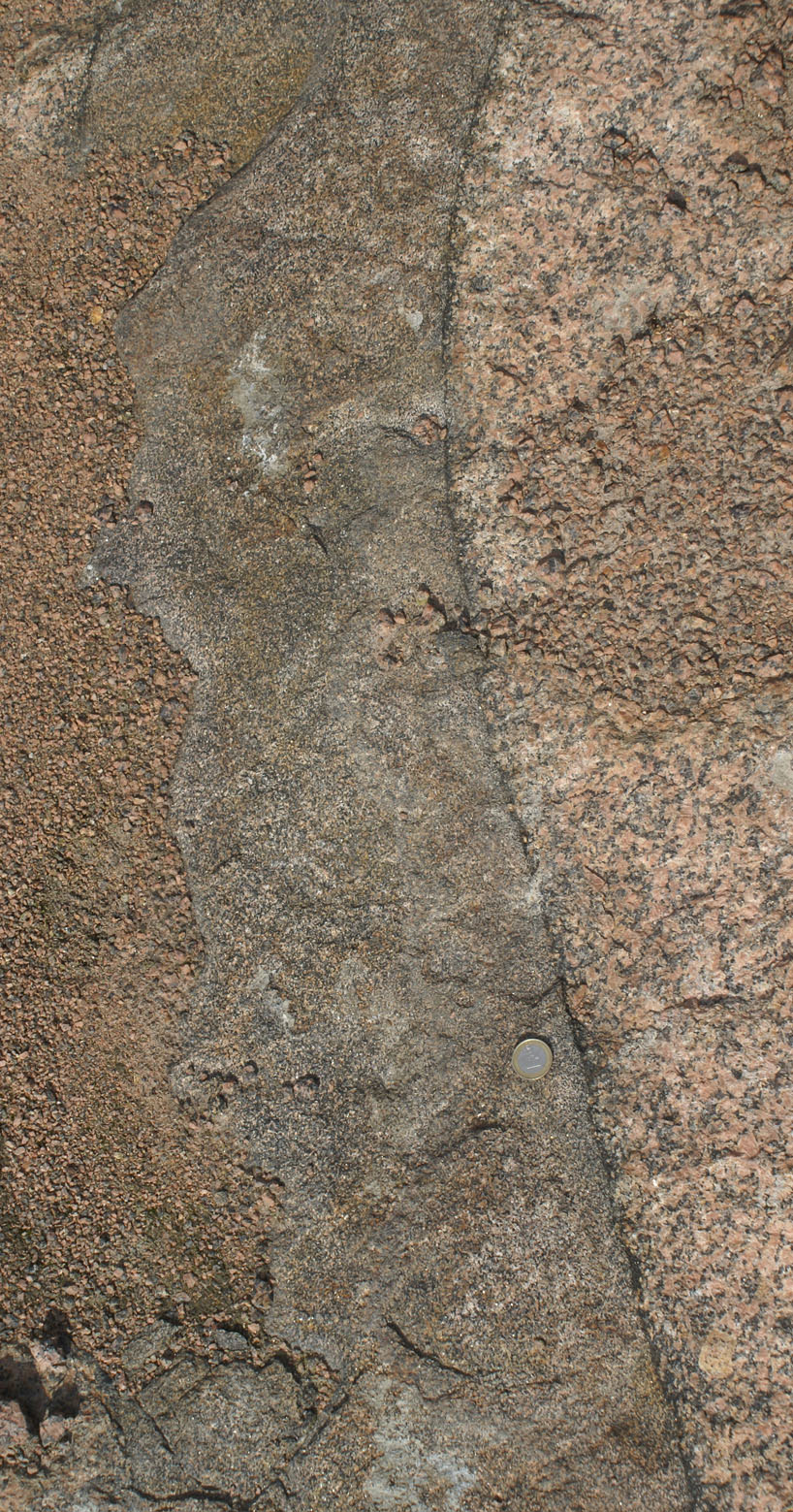
| Figure 5a) Ptygmatic fold with well-defined folded margins merging continuously with a diffuse leucocratic patch elongated parallel to the main foliation. | Figure 5b) Ptygmatic fold with well-defined folded margins becoming diffuse to the left. | Figure 5c) Same. This pattern is interpreted as a disrupted dykelet by local increase in melt fraction in surroundings during of after dyke propagation. Due to the intense folding of the veinlets and the axial planar orientation of the diffuse melt patches, I interpret the pattern to result from folding during melting. |
 |
 |
|
Figure 6a) Ptygmatic fold with
well-defined folded margins merging continuously with a diffuse leucocratic patch elongated parallel to the main foliation. |
Figure 6b) |
c) Migmatites from Spikarna (N6633592, E3287503) in Sederholm 1907: melt seggregation and extraction from migmatitzed pre-orogenic supracrustals and synorogenic tonalties
 |
 |
 |
| Figure 7a) Svecofennian supracrustal rocks which underwent partial melting. Leucosomes in boudin necks (upper left corner). Leucosomes accommodating strain in fold hinges in mafic layer in lower right corner. | Figure 7b) Detail of a) showing two triangular-shaped leucosomes in fold hinge. The upper one (upper right) wedgest out to the right and becomes increasingly leucocratic and looses early gneissic fabric, suggesting the filtering out of solid particlers (filter pressing). The lower one (lower left), wedges out to the left. | Figure 7c) Detail of b), notice funneling and merging of leucosomes, becoming increasingly magmatic and leucocratic to the right. |
|
|
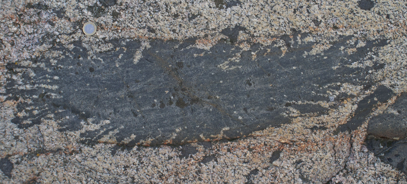
| Figure 7d) Detail of a). Leucosome, indicative of melt segregation, in boudin necks in stromatic migmatite. Notice dykelet in the upper left linked to boudin leucosome, suggesting a escape path for magma accumulated in the neck. | Figure 7e) Leucosome on the right in d) cutting across but linked continuously with layer-parallel leucocomes in the stromatic migmatite. On the left-hand-side it is associated with a fold, close to and immediately right of the coin it is associated with a cuspate fold pointing to the right, which becoms a gentle monocline further to the right. Relationships in d) suggest that the structure represents a boudin neck that later slipped slightly. |
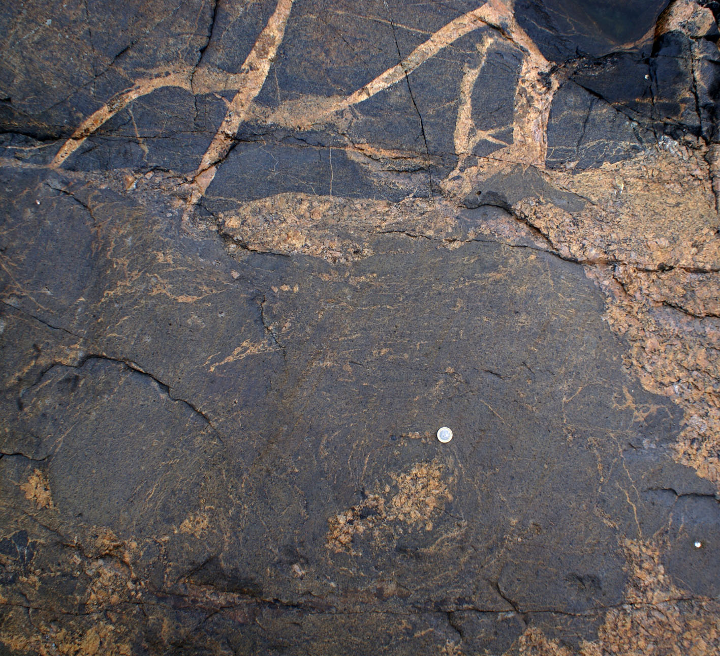
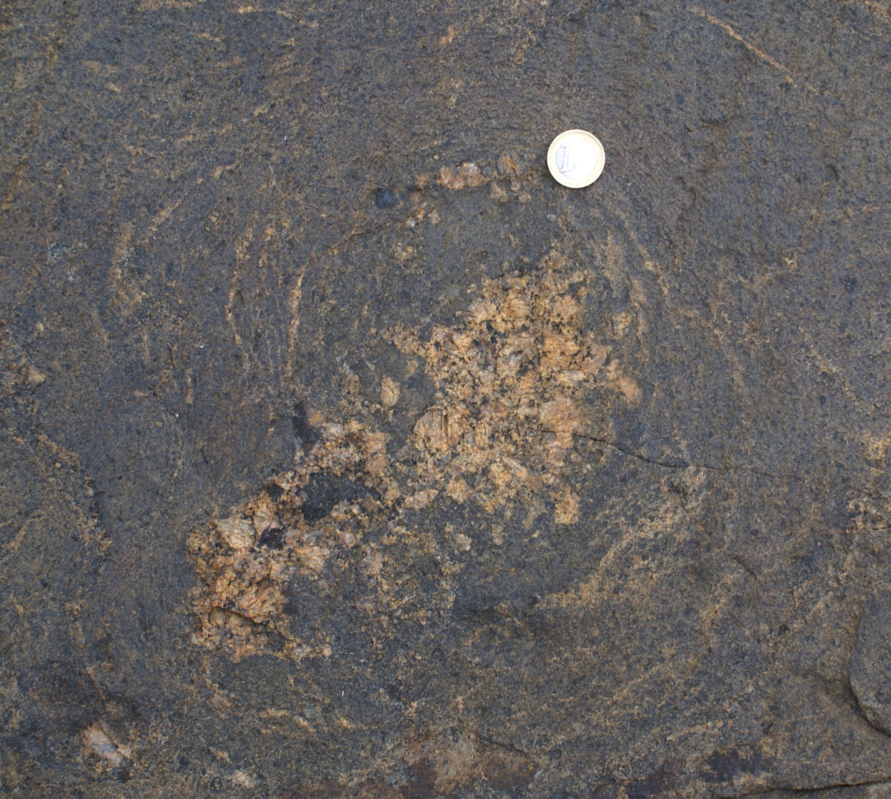
d) Magma Mingling
|
|
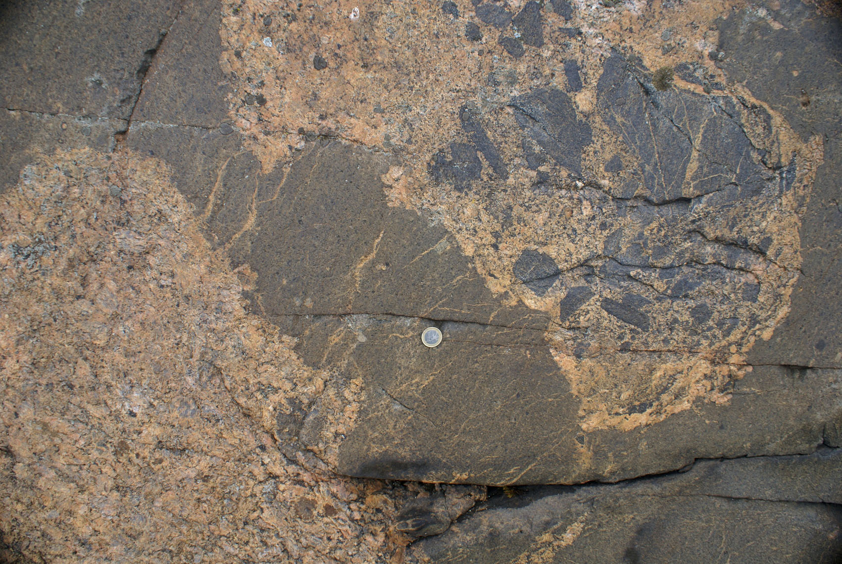
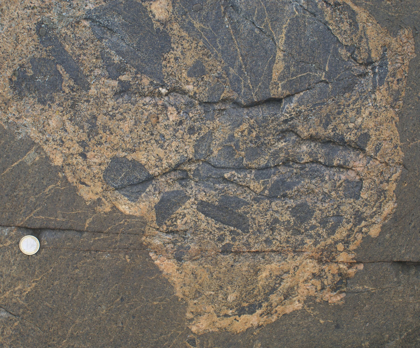
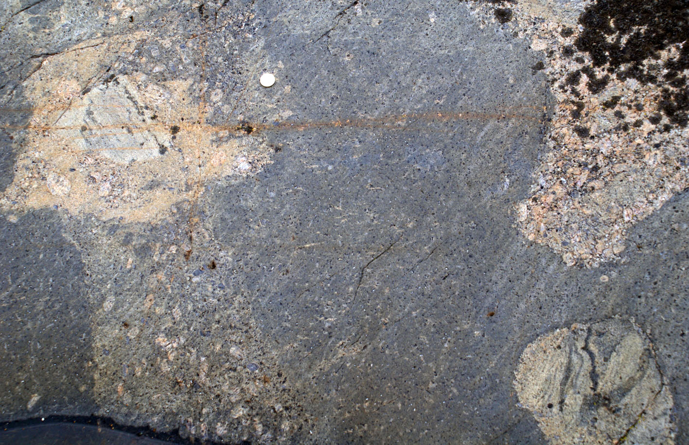
| Figure 8a and b). Åva harbour. Contrast in behaviour of felsic magma intruding mafic rocks. In the upper part of the photograph, felsic magma intrudes a solid mafic xenolith forming pegmatitic granite dykelets linked with finer grained granite outside the xenolith. In the lower part, mafic and felsic magmas mingled intensely as they rotated together now forming a swirling pattern marked by narrow circum-centric felsic veinlets in mafic surroundings and a core of coarse granite (best seen in detail beside). |
|
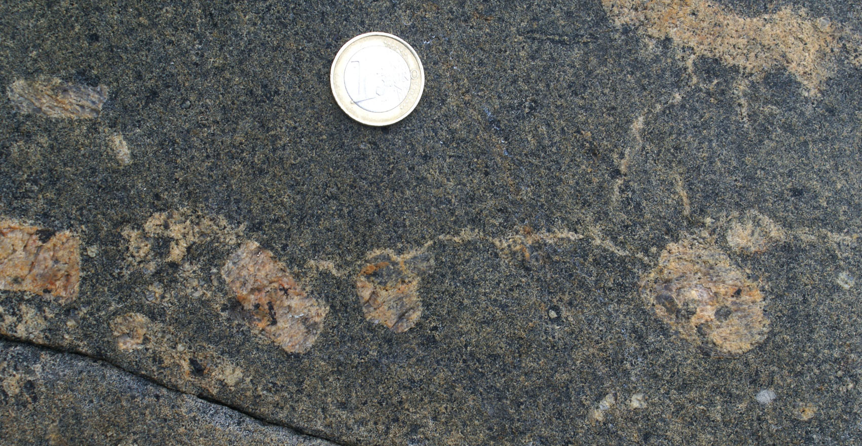 |
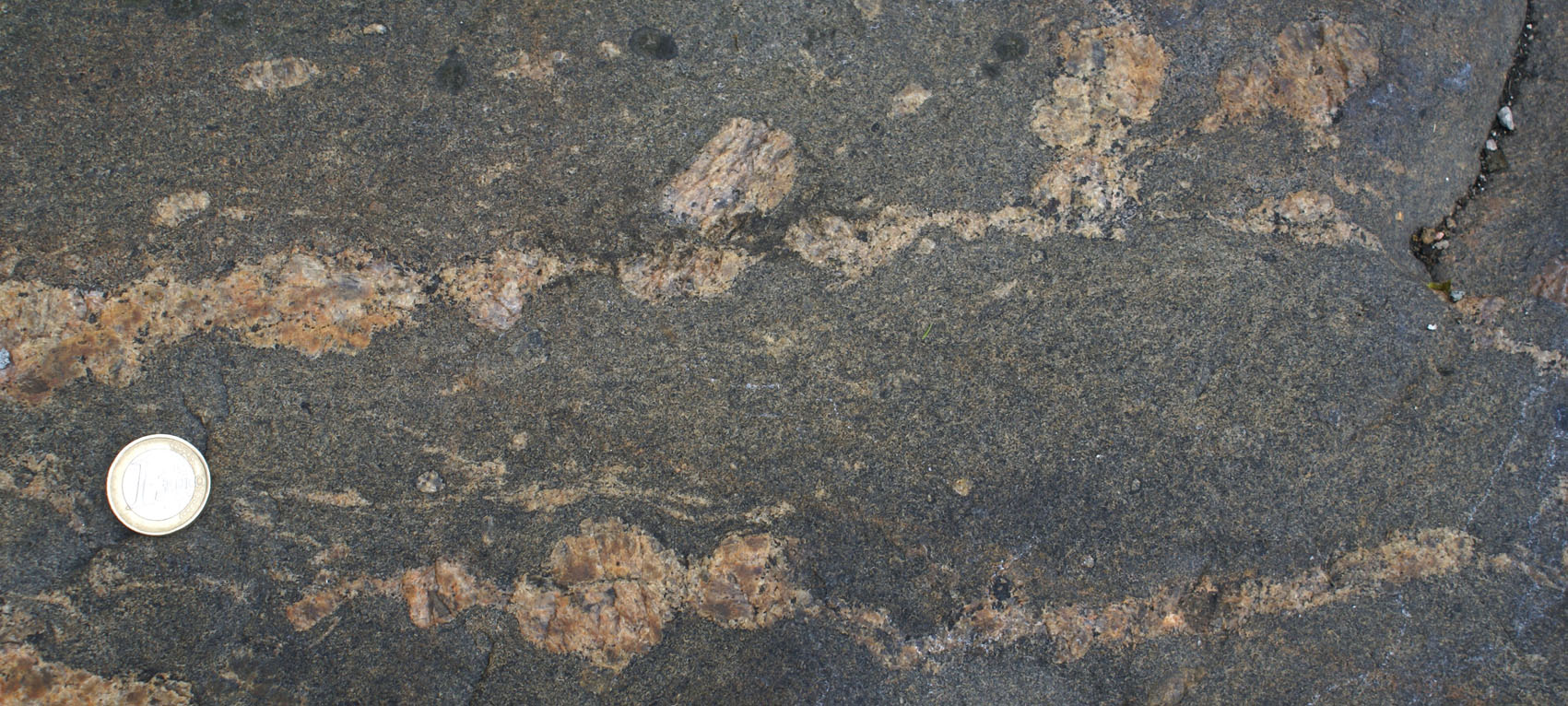
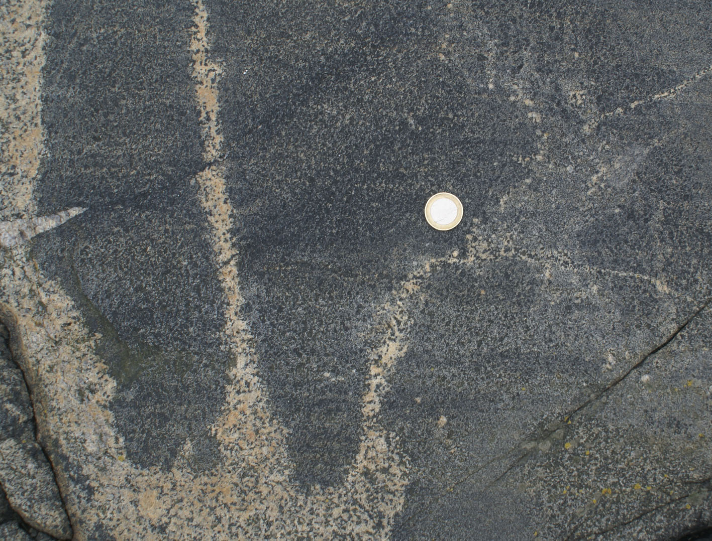
| Figure 9a) Åva harbour. Granitic veinlets linking K-feldspar phenocrysts in monzodiorite. Result of magma mingling and squeezing of interstitial granitic melt and filtering of phenocrysts. | Figure 9b) Same as a) in detail. |
 |
 |
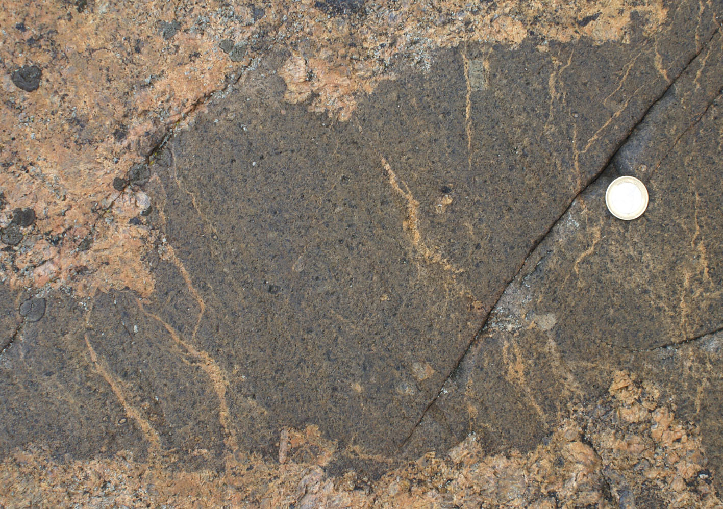
| Figure 9c) Northern part of Enklinge island at Vindarskär ("the outcropt that looks like a cow" student quote, Stop 14 in guide book N6709573, E3161919). Different stages of mafic pillow disaggregation in granite | Figure 9d) |
|
 |
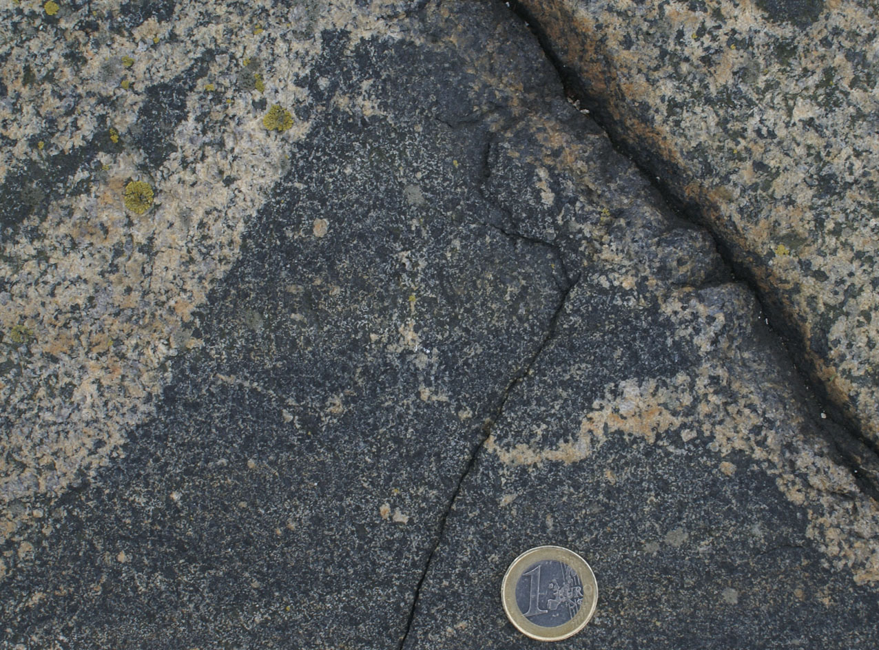
| Figure 10a) Northern part of Enklinge island at Vindarskär (stop 14 in guidebook). Gradation from granite indenting a pillow in the lower left to isolated feldspar phenocrysts becoming more sparse to the upper right of the pillow. | Figure 10b) See Kelemen's experiments. |
 |
|
 |
| Figure 11a) Åva harbour. Minlging of granite and monzonite of the Åva intrusion, and brecciation of xenolith inside granite. | Figure 11b) Detail of brecciated xenolith inside a pool of granitic rock mingled with surrounding monzonite. Notice the extent of the brecciation, forming clasts smaller than 0.5 cm (in 2D) | Figure 11c) Irregular veinlets of granite intruding the monzonite band separating the two pools of granite. Notice that similar to patch migmatites, veinlet walls become diffuse and merge with the matrix of the monzonite forming leucocratic patches. |
|
 Figure
12b) Same as a) with a patch of leucocratic monzonite on the
right.
Figure
12b) Same as a) with a patch of leucocratic monzonite on the
right. |
| Figure 12a) Stop between Bränd;öharum and Påvskär. Mingling between granite and monzonite. Notice how the tip of the intruding granite veinlet on the right-hand-side becomes diffuse forming a leucocratic patch in the monzonite akin to a patch migmatite, but in this case resulting from melt inflow rather than outflow. |
e) Ductile Fractures
|
|
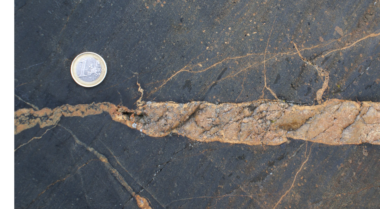
| Figure 13a) Lil-Lappo (N6709753, E3161919). Irregular, zig-zagging tonalite-filled fractures in two different orientations in hornblende-cumulate, possibly formed as a result of segregation of interstitial melt. | Figure 13b). |
|
 |
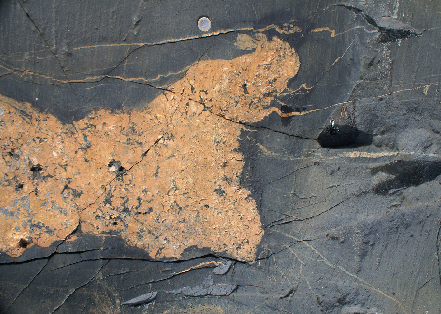
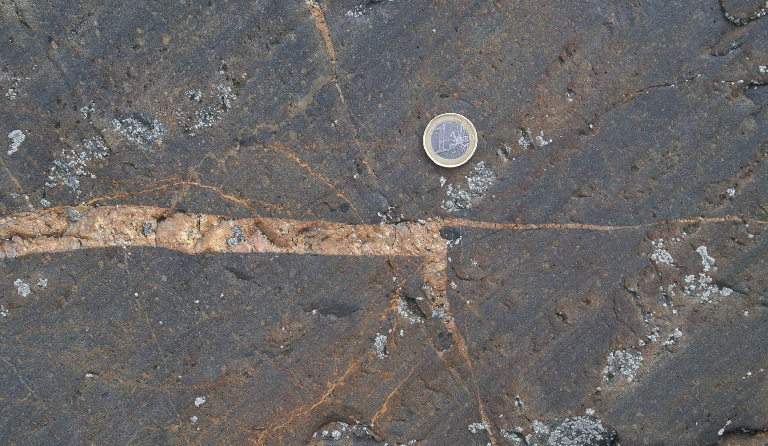
| Figure 14a) Stop between Brändöharum and Påvskär. Magma mingling resulting in irregular fractures repeatedly splitting (left-hand side). | Figure 14b) Stop between Brändöharum and Påvskär. Right-angle fractures filled with granite in monzonite. |
 |
 |
 |
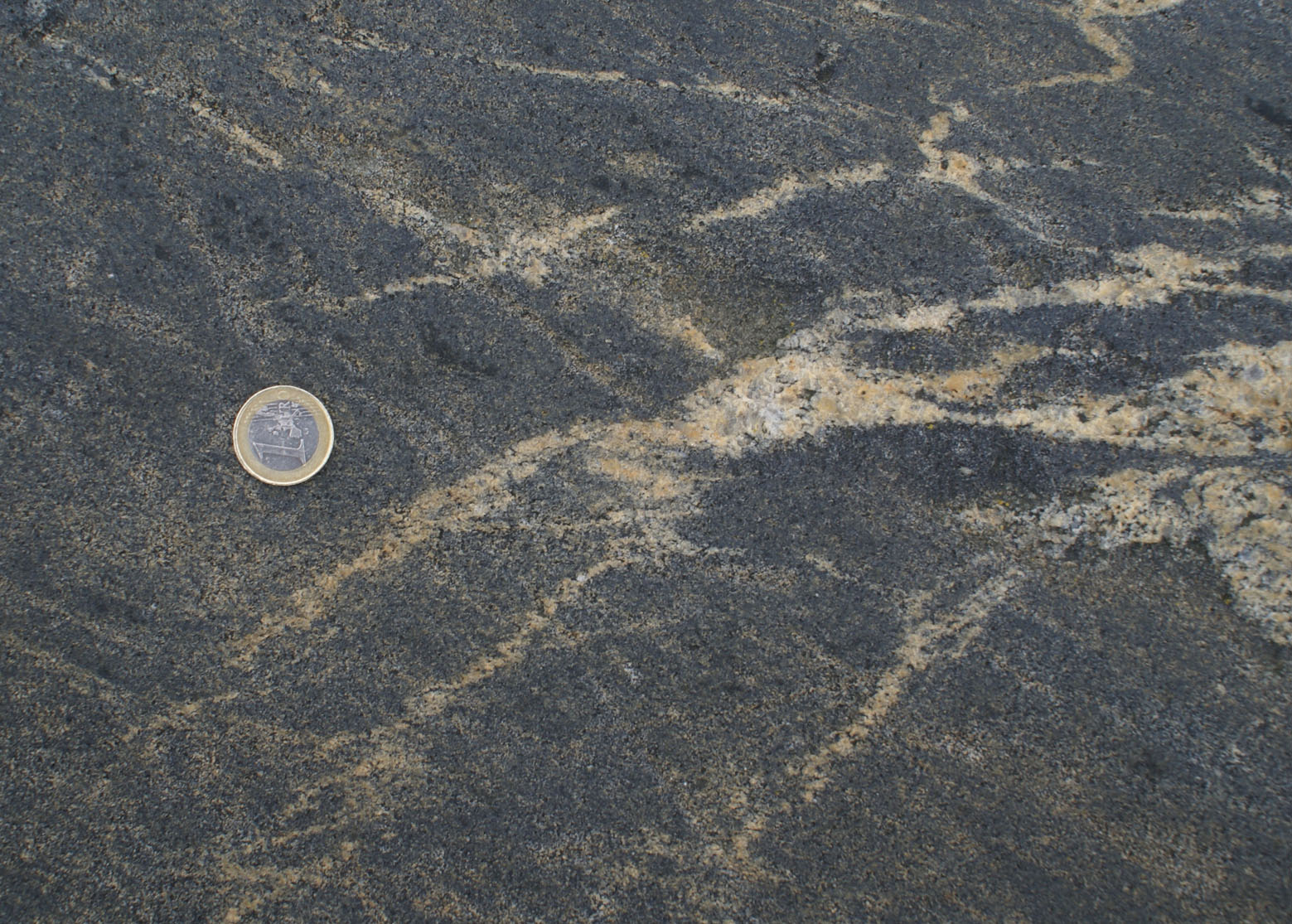
| Figure 15a) Lövgrund (stop 6, N6650561, E333240). Ductile fractures in migmatite following mode II fractures in "pre-orogenic mafic and felsic volcanics with carbonate-rich interlayers cut by synorogenic gabbro-tonalite dykes. All rocks are later folded and migmatized." | Figure 15b) Enklinge island. | Figure 15c) Fractures related to a leucocratic accumulation. |
f) Partial Melting at Dyke Margin in Peperite, Enklinge island (Stop 12 in guidebook, N6706142, E3155693)
 |
 |
| Figure 16a) Mafic dyke intruding peperite. During metamorphism the region close to the altered margin of the peperite (darker vertical band) undergoes melting and melt intrudes the dyke to the right. | Figure 16b) Detail of a) showing a decrease in mafic minerals from left to right, and irregular dykelets propagating outwards from the main dyke. |
 |
 |
| Figure 16c) Inside a mafic dyke, pegmatitic dyke, presumably formed by melting at the margin and intruding into the dyke (as inferred in 16a, b). Note also irregular dykelets propagating from it. | Figure 16d) Tip of felsic dyke in mafic dyke intruding peperite. A box-shaped tip splits into two narrower dykes are right angles to each other. |
g) Brecciation and assimilation of calc-silicate roof pendant in the central part of the Åva intrusion (Stop 19 Nottholm N6720892, E3173346)
 |
 |
 |
| Figure 17a) Breakup of roof pendant. | Figure 17b) Breakup of roof pendant. | Figure 17c) Assimilation of calc-silicate roof pendant in granite. |
|
 |
 |
| Figure 17d) Hybrid monzonite with calc-silicate enclaves, partly assimilated. | Figure 17e) Detail of d. Granite mingled with monzonite. In monzonite two football-sized enclaves: the one on the left is an altered/assimilated calc-silicate(?) as evidenced by partly preserved layering; the other is granitic. | Figure 17f) Granitic dyke intrudes calc-silicate across layering and then along layering assimilates it and new, larger amphiboles form. |
h) Ladder dyke.
(N67..., E31...)

|
 |
| Figure 18a) Ladder dyke on vertical wall. Notice the very wide mafic accumulation at the base. | Figure 18b) Mafic accumulation in granite defining a cone. It has a sharp outer margin and grades inwards to the typical granite. |
|
 |
| Figure 18c) Ladder dyke on horizontal plane. More mafic and finer grained than surrounding rocks. | Figure 18d) Margins of ladder dyke on horizontal plane (dyke above, country rock granite below). Hornblende accumulations on the external side of the dyke. |