 |
Magma Migration, Folding and Disaggregation of Migmatites the in the Karakoram Shear Zone, Ladakh, NW IndiaRoberto Weinberg, Monash University, Australia Geordie Mark, Monash University, Australia |
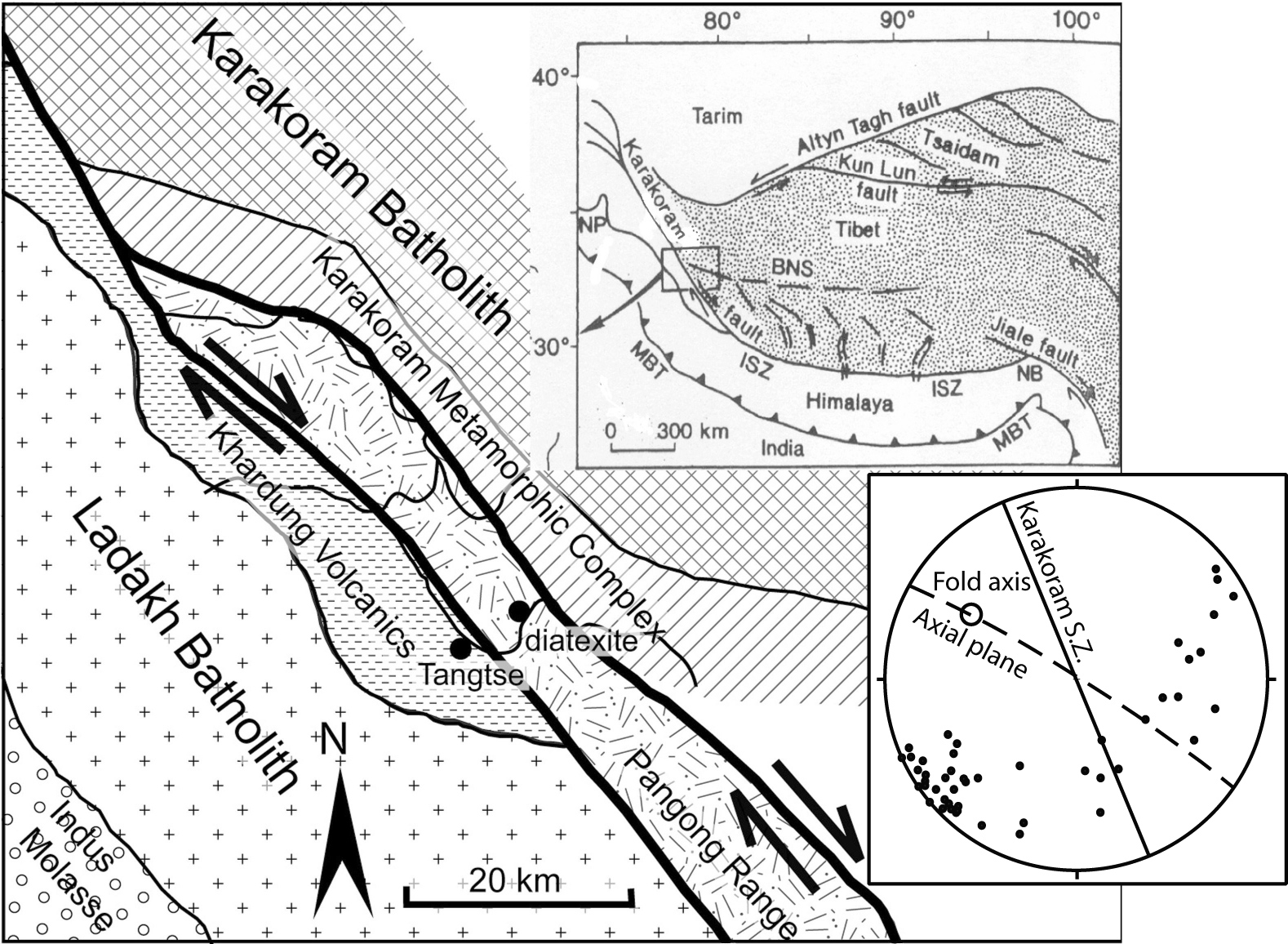
| Figure 1. Geological map of the are surrounding the Karakoram Shear Zone in NW Ladakh, India. The Ladakh Batholith and Khardung Volcanics represent an early, pre-collisional calc-alkaline sequence. The diatexite layer studied in this paper is exposed in the gorge transecting the Pangong Range and its Pangong Metamorphic Complex, which is bound on either side by strands of the Shear Zone. The Karakoram Metamorphic Complex and the Karakoram Batholith crop out to the NE. Insert: stereonet defining NW-trending upright folds within the broader 140-trending Karakoram Shear zone. |
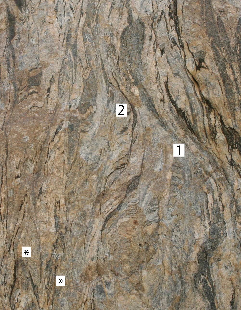
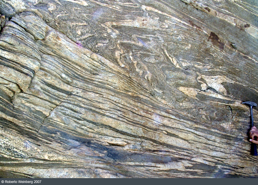
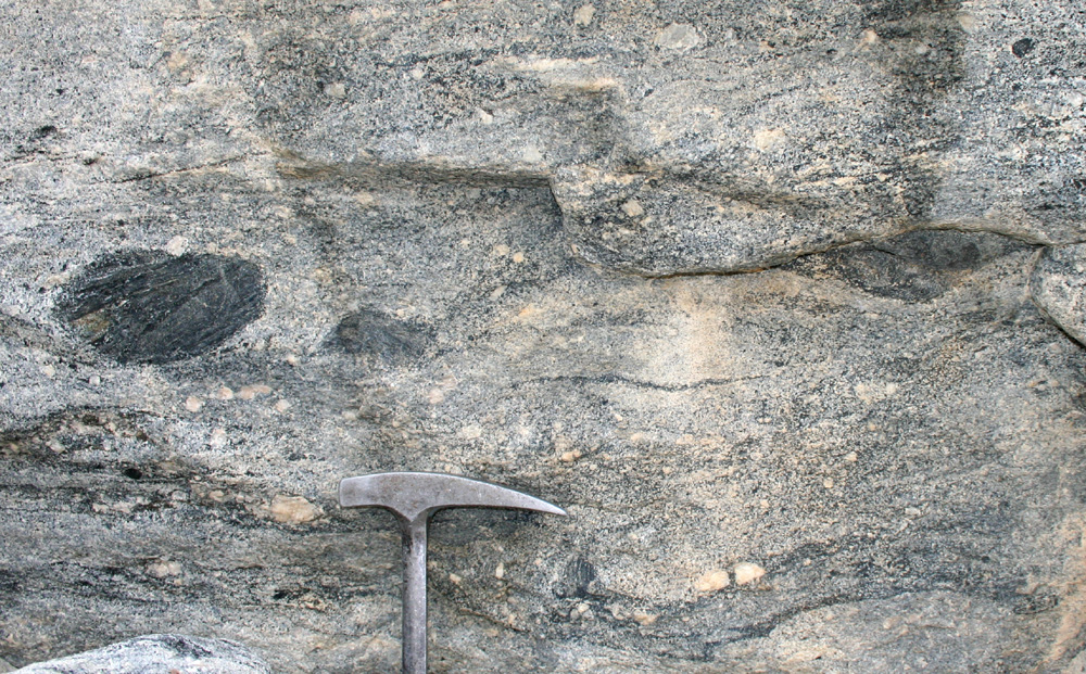
| Figure 2. Vertical outcrop of hornblende-bearing sequence within the diatexite zone in Fig. 2, showing advanced stages of transposition where folds are tight (*) and limbs nearly transposed (1). Layers in metatexite defining asymmetric folds are dragged upwards into a transposed zone (2). Note antiformal fold closures (*) and absence of synformal closures. Scale: vertical side of image is 1.5 m. |

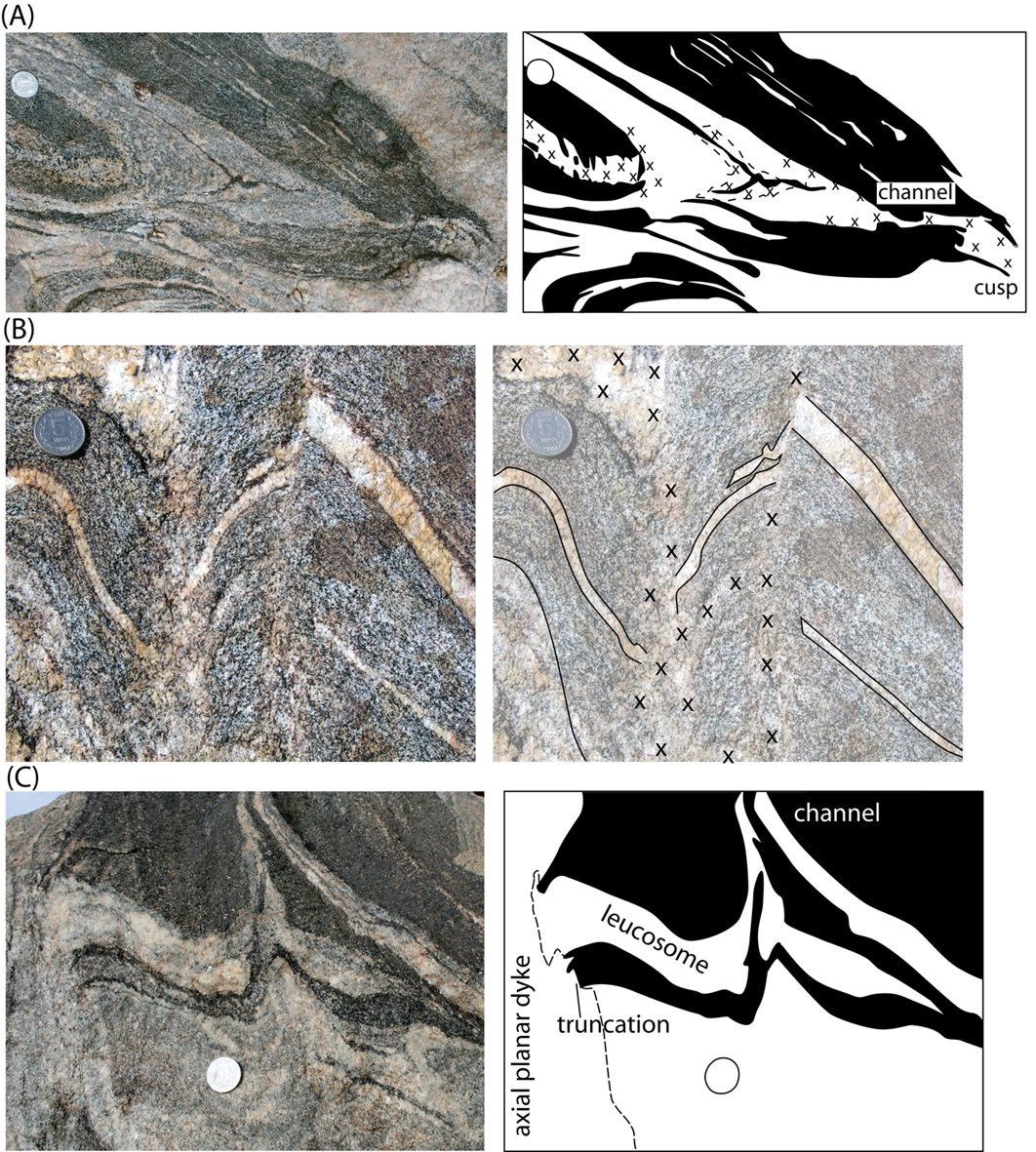
| Figure 3. (A) Summary of the main features of magma-assisted fold transposition in Tangtse. Lenses with different tones of grey and in black represent schematically melanosomes and mesosomes. 1. Layer truncation across cuspate hinge zone, with or without axial planar leucosome. Cusps point outwards from the fold core. 2. Limbs are defined by asymmetric "parasitic fold blocks" bound by well-developed axial-planar foliations. Inside each parasitic fold block, folds are disharmonic with varying profiles and degrees of rotation towards the axial planar orientation. Parasitic fold blocks can be found in fully transposed parts of the diatexite as remnants of the transposition process (2'). 3. Axial planar foliation acted as shear planes accommodating considerable fold stretching. Such foliations may be filled with mafic residual material (melanosome), leucocratic material (leucosome), or mesocratic material. Layers may be continuous across axial planar foliation (left of 3 in figure), or may be truncated and discontinuous (right of 3). 4. Leucogranite dike trends diagonally across the fold from outer parts of limbs towards the fold closure in a manner broadly conformable with the shape of the fold. This is achieved by axial planar leucosomes side-stepping inwards through layer-parallel leucosomes. 5. Mafic minerals are dragged from melanocratic layers into the axial planar leucosomes and dikes forming schlieren. 6. Parasitic fold blocks undergo increasing rotation towards the transposition plane, outwards from the hinge zone (arrow labelled with "rotation"). 7. Fully transposed regions are characterized by schlieric diatexites containing remnant structures of the transposition process, such as an individual asymmetric fold blocks (2'), and rootless folds (8). |
| Figure 4. Some typical features associated with metatexite folds. (A) Leucosome in fold hinge zone, cutting across mafic layers forming a cuspate hinge on the right, defined by dragged and reoriented biotite and hornblende grains. Leucosomes are diffuse in the gneissic layer and more focused in mafic layers. Melt is interpreted to have moved to the right, from the fold core on the left, outwards, forming a melt channel through the less permeable mafic layer which is disaggregated the cuspate fold tip. (B) Irregular axial-planar leucosomes with poorly-defined margins in fold hinge zones (marked with x in line drawing) merging continuously with layer-parallel leucosomes with sharp margins and narrow (mm wide) melanosome rims. Axial-planar leucosomes are interpreted to represent melt extraction paths which have partly drained layer-parallel leucosomes causing disruption and truncation. (C) Metatexite truncated on the left-hand-side by a grey axial planar dike with diffuse internal banding parallel to its margin. Leucosome and melanosome layers fold into a narrow channel through a biotite amphibolite layer. Melanosome layer is incipiently disrupted by the process. All images from blocks. |

| Figure 5. Cuspate hinge zones: (A) a tight, cuspate hinge zone is linked to a pegmatite body (close to hammer) in contact with broken and rotated parts of a competent amphibolite layer. The sharp cuspate fold hinge, the presence of pegmatite and the break-up of the amphibolite layer suggest that melt once drained through the hinge zone (see Fig. 5a) and that the fold cusps point towards the direction of melt flow. Pegmatite accumulation on one side of the amphibolite suggests ponding before amphibolite disaggregation. (B) Dragging of melanosomes and leucosomes towards a fold hinge that cuts across a competent amphibolite layer on the right-hand side, interpreted to be a melt channel. Melanosome bands merge into the cuspate hinge zone indicating loss of leucosome material. Note melanosomes are not continuous layers, but thin out laterally. (C) Fold with cuspate hinge zone and well-developed, leucosome-free axial planar foliation. Layers are abruptly truncated or change width across well-developed axial planar foliations. For example, the coarse-grained, hornblende-rich melanocratic layer marked m1 in line drawing (lower part of image) is discontinuous across an axial planar foliation marked by hornblende grains spread along its length (Hbl on line drawing). Similarly, the mesocratic layer to the right of the coin changes width abruptly across the axial planar foliation. The leucosome layer in upper right is truncated by the axial planar foliation, the other side of which there is instead a narrow melanosome (m3). Combined these features suggest that layer-parallel leucosomes and melanosomes were extracted along the axial planar foliation, which are inferred to have acted as a melt conduit. All images from loose blocks. |
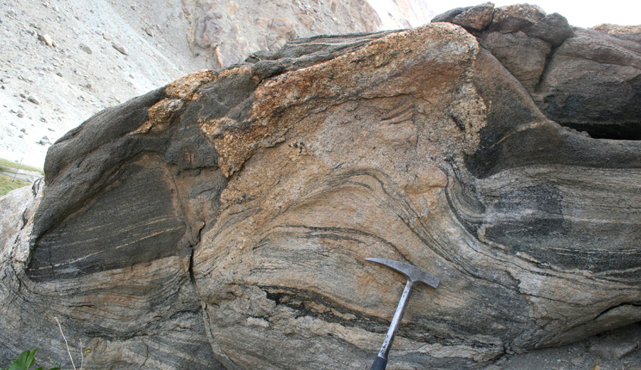 Figure 6. Pegmatite accumulation on the concave side of a gently folded contact between metatexitic migmatite and amphibolite layer. Pegmatite intrudes amphibolite above and has diffuse contacts with migmatite below. Higher amplitude of pegmatite intrusion compared to that of the ghost stratigraphy in the migmatite-pegmatite transition zone (close to hammer), indicates pegmatite drainage from the migmatites. |
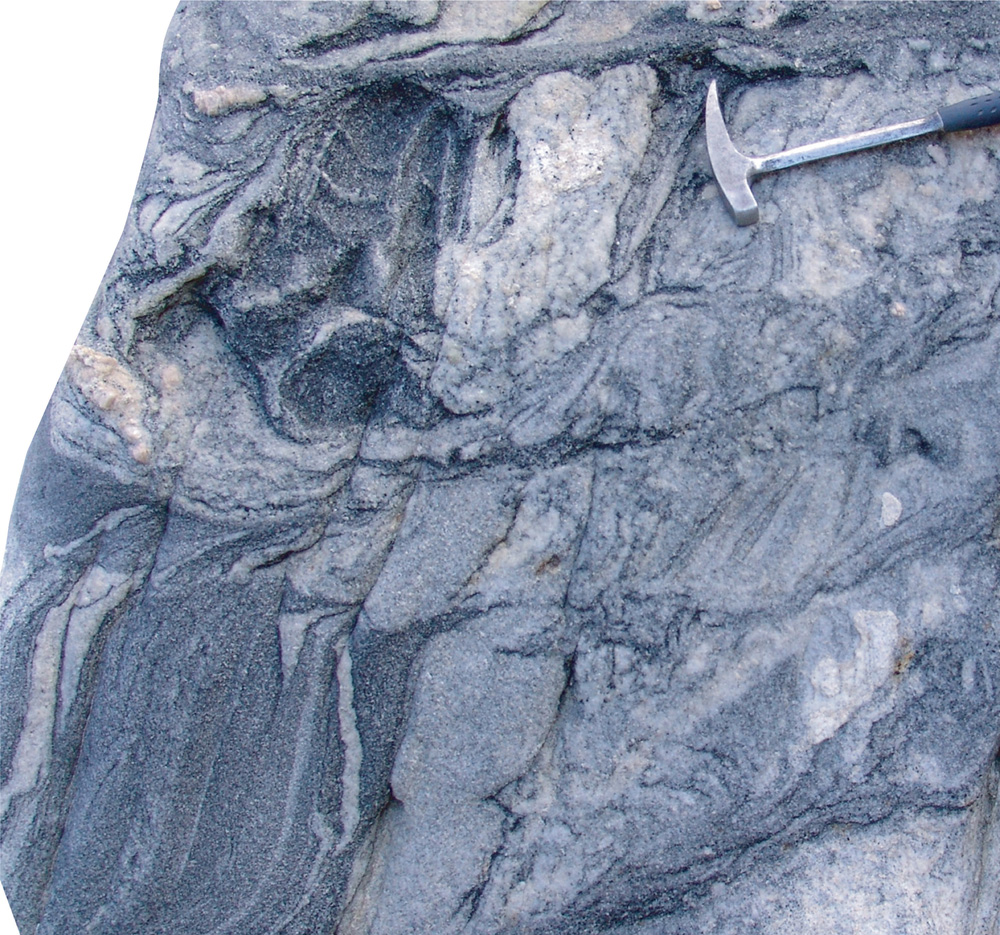 Figure 7. Leucosome cuts through, drags and disaggregates layering. Schlieren cusps and increased disaggregation suggest leucosome flow to the right. Features are exaggerated by cut effect due to block surface being at 40o to migmatite layering. |
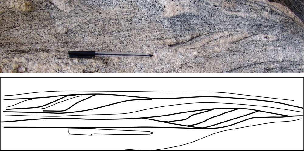
 Figure 8. Gradual transposition of layers towards a hinge zone. Parasitic fold wavelength decreases and amplitude increases towards the transposed zone as a result of stretching and rotation of blocks. Ultimately, layers are truncated by the 10 cm-wide leucosome band with biotite-hornblende schlieren (lower-central part of figure), interpreted to be a channel-way through which melt escaped, transposing layering caught up in the flow. |
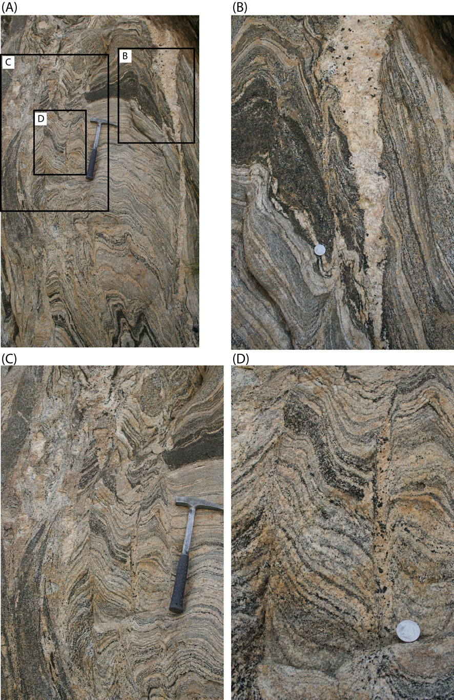
| Figure 9. Overview and details of a steep exposure recording many features of the transposition process. (A) Metatexite fold preserved inside a generally transposed diatexite (left-hand side of image). Transposed layering strikes 120o and dips nearly vertically and layering in metatexite is at high angle to transposition plane. (B) Detail of leucosome in the axial plane of a cuspate fold merging upwards with a larger dike with euhedral porphyroblasts of hornblende (upper right-hand-side). This dike is roughly parallel to the axial plane of the fold and widens upwards. (C) Detail of transposed layering on left, and angular, cuspate folds on right, truncated along the hinge zone by axial planar foliation. Note truncation of the thick mafic layer above hammer by axial planar leucosome and fold train. Note also that these fold trains get disrupted and truncated by pegmatitic granite further up. Fold profiles just left of the hammer change from close angular in the upper part, to increasingly open downwards (below hammer). (D) Detail of angular cuspate folds in (C) showing both layer truncation and continuity of layers across the axial planes with hornblende-bearing leucosome. |

| Figure 10. (A) Axial planar foliation in folded metatexite. Folds are disharmonic and limb in the upper part of image is strongly stretched and rotated towards the axial planar orientation. Note pen close to the centre as scale. (B) Detail of A, layers truncated by axial planar foliation, and angular fold with diffuse accumulation of leucocratic material along the hinge zone. Layers dip 50o into exposed surface. |

| Figure 11.(A) Metatexite preserving primary banding at high angle to the axial plane of folds, which act as shear zones, truncating layering (e.g. amphibolite layer in upper left). Hammer for scale in bottom right. Box shows the position of B. (B) Detail of disharmonic folds and layer truncation. Axial planar band contains partly disaggregated melanocratic bands and schlieren in granitic material. Layers in the upper half of the image are unrelated to those in the lower half. The melanocratic layer m1 thins to 1/30 of its width in the fold in the middle of the image, and layer m3 thins to 1/10 of its width in the hinge zone. These features suggest bulk mass loss, possibly recorded by disaggregated melanocratic bands in axial planar transposed zones. |
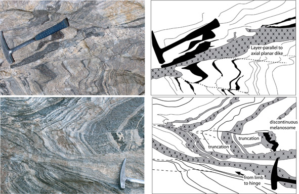
| Figure 12. (A) Hornblende leucogranite crosses a fold limb alternating between conforming to folded layering and to the axial planar foliation, causing it to trend diagonally towards the hinge zone. (B) Axial planar leucosome (under hammer) truncating fold limb and stepping inwards towards hinge zone. This leucosome is linked continuously with layer-parallel leucosomes (grey shading with crosses in line drawing). Fold profile changes from an open fold on the right into a sharp acute fold on the left. Layers on the lower limb (left of hammer) are strongly rotated towards the axial planar orientation. Note layer mismatch across the hinge line of the open fold and discontinuous melanosome (black in line drawing) in centre-right of the image. |
 (A) |
 (B) |
| Figure 13. (A) Advanced stages of disaggregation forming a diatexite including numerous blocks of refractory material and grains, as well as schlieren and bands of different composition. (B) Diatexites preserve also parasitic fold trains recording late stages of transposition. |