Leucosomes from Malung Thokpo, Zanskar Mountains, High Himalaya
|
Leucosomes from Malung Thokpo, Zanskar Mountains, High Himalaya
|
|
|
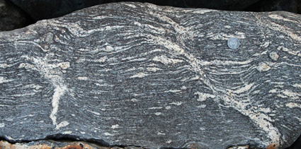
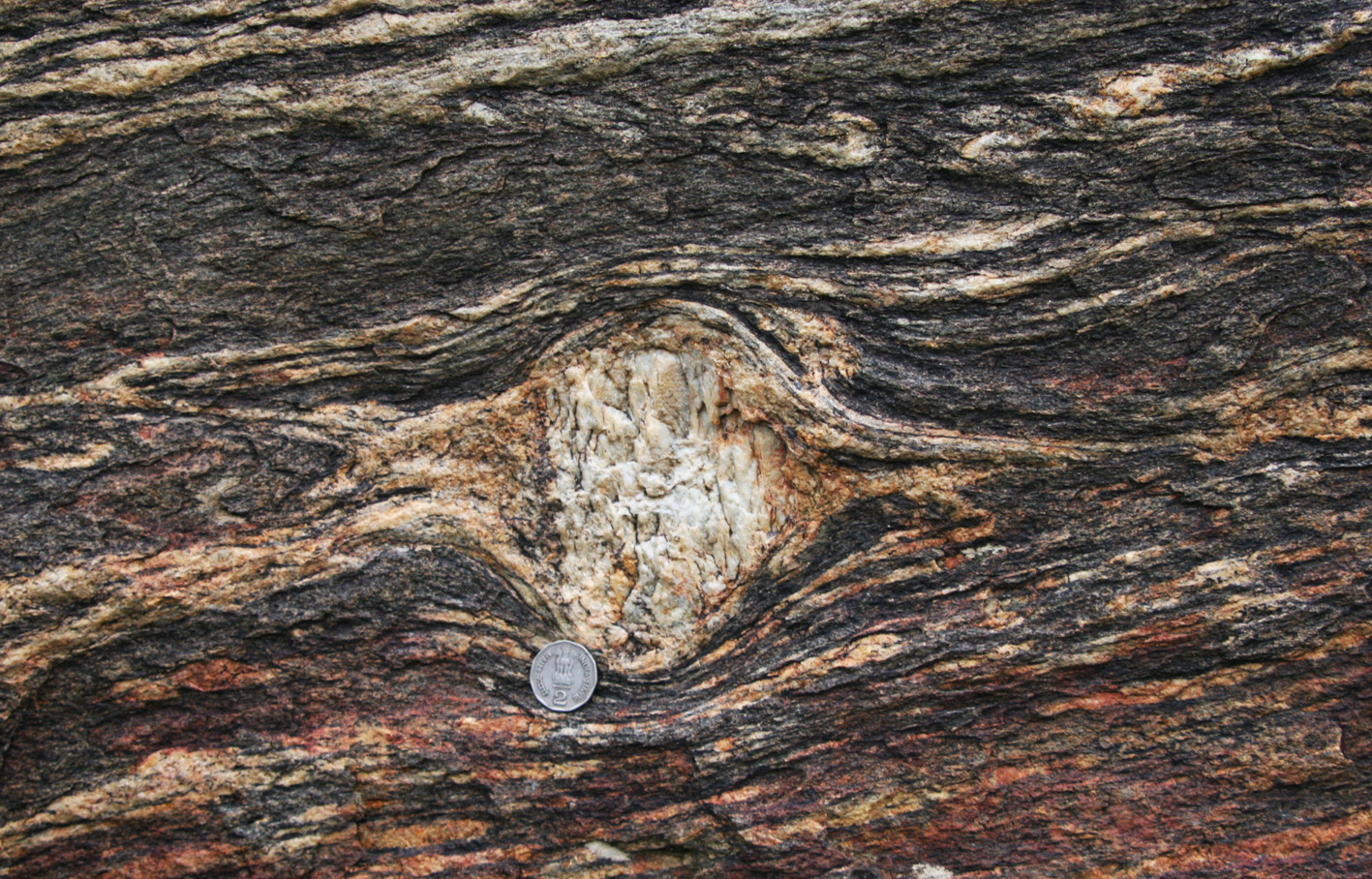
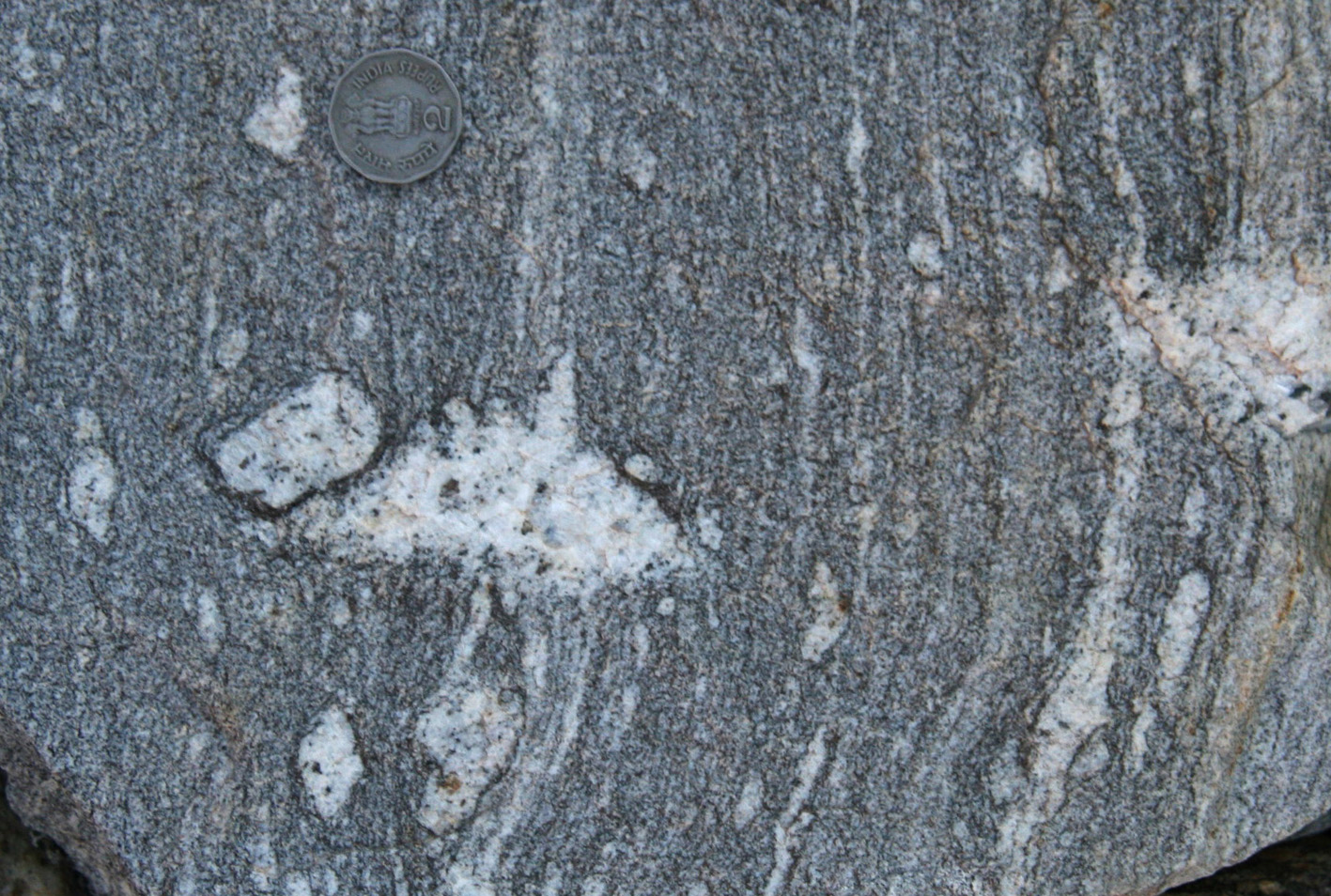
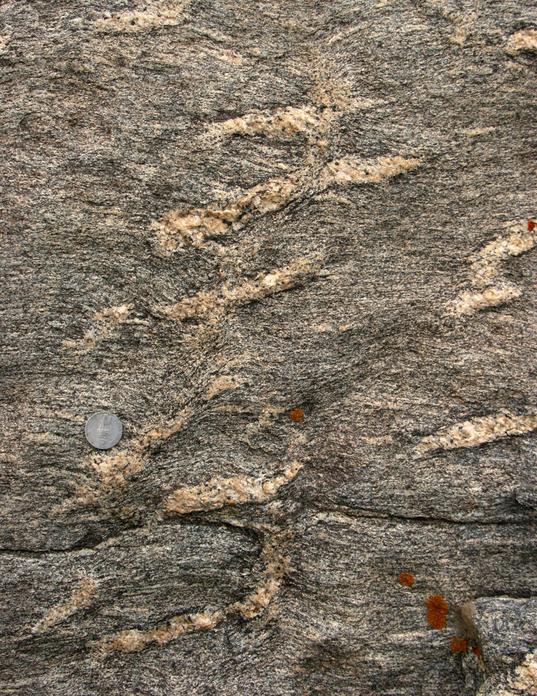
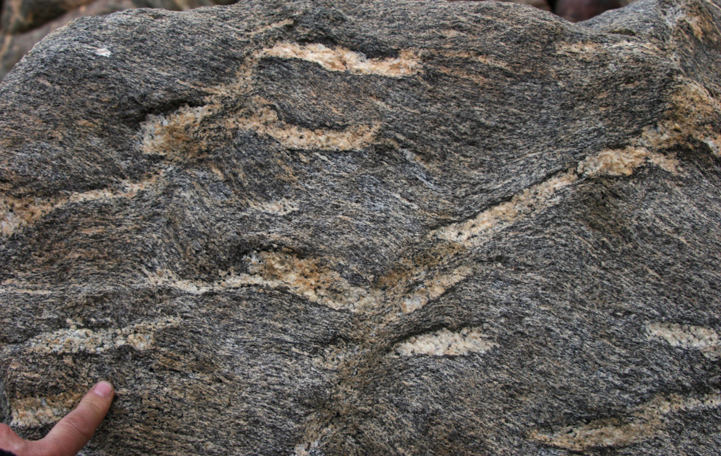
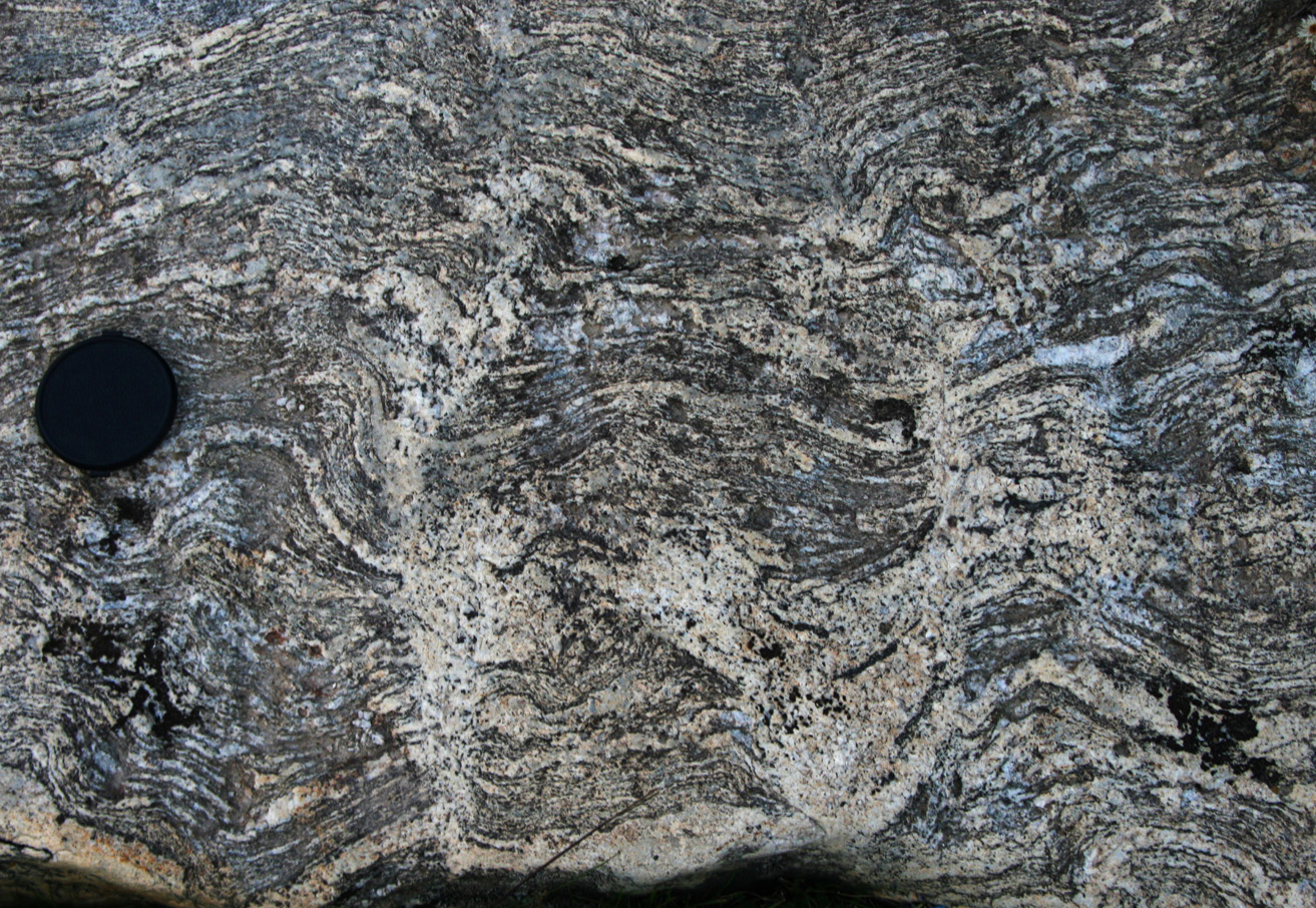
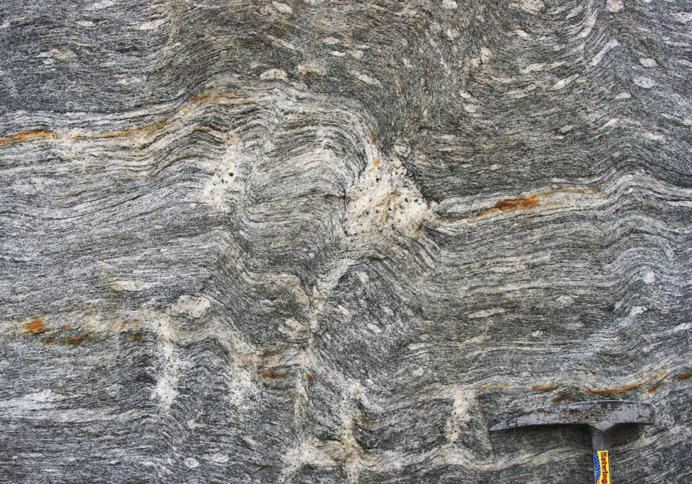
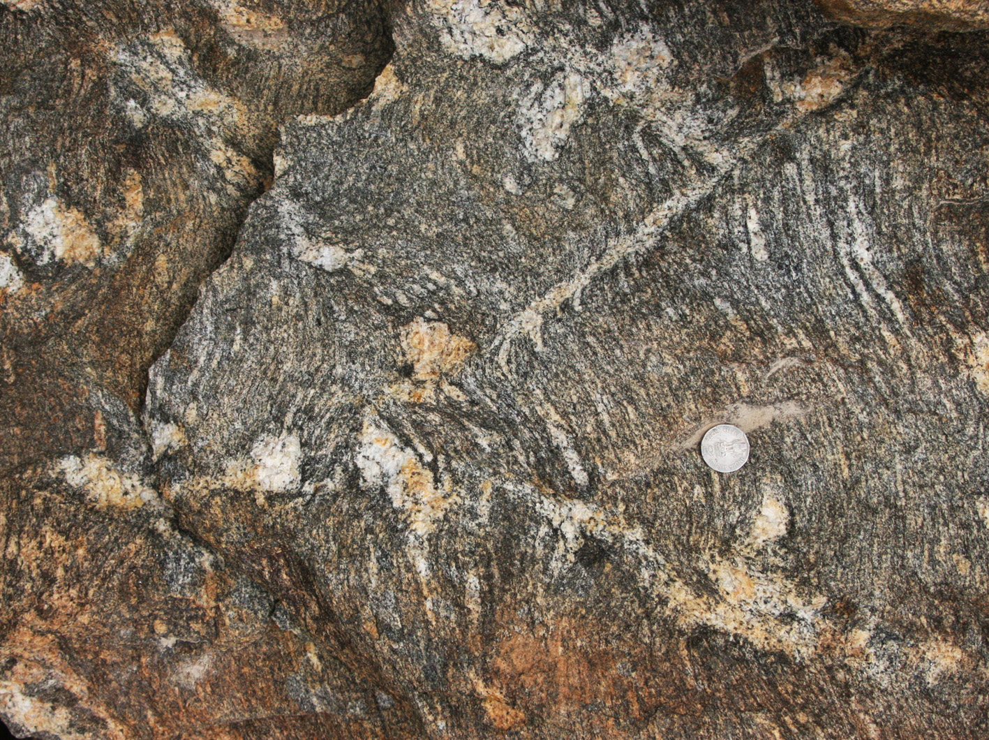
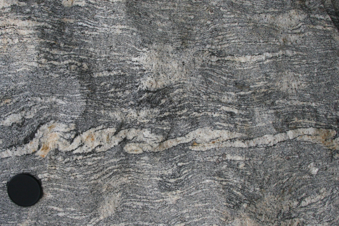
| a) K-feldspar porphyroclast with asymmetric tails of leucosomes indicative of top to the left. Interpretation: normal shearing during melting (water-fluxed melting, no peritectic minerals). Note truncation of layering across a diagonal plane (from lower right to upper left) indicative of movement along that plane. Photo parallel to lineation and perpendicular to foliation. | b) Example of K-feldspar porphyroclast with asymmetric leucosome tails indicative of top to the left shearing in the presence of melt. Photo parallel to lineation and perpendicular to foliation. |
|
|
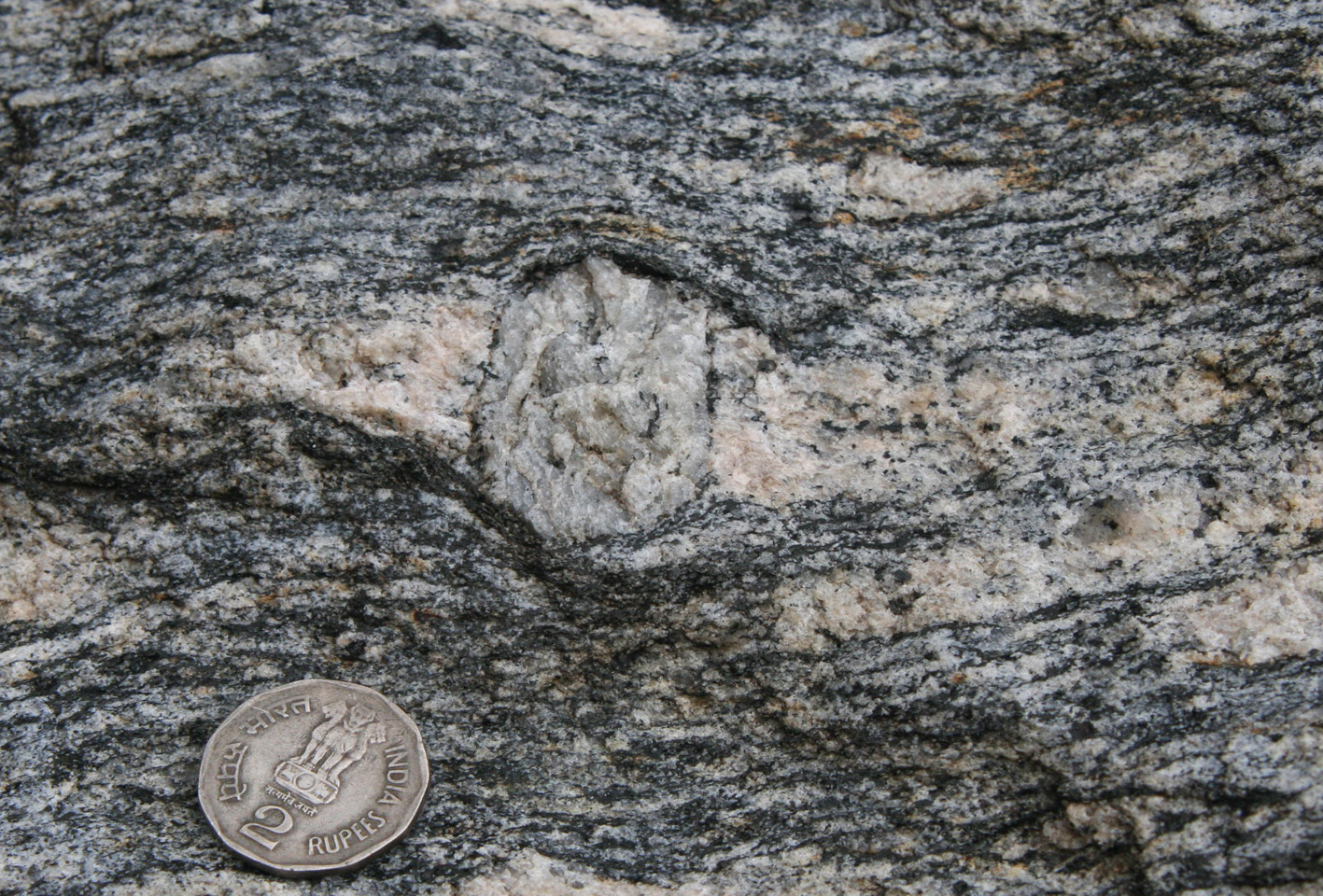
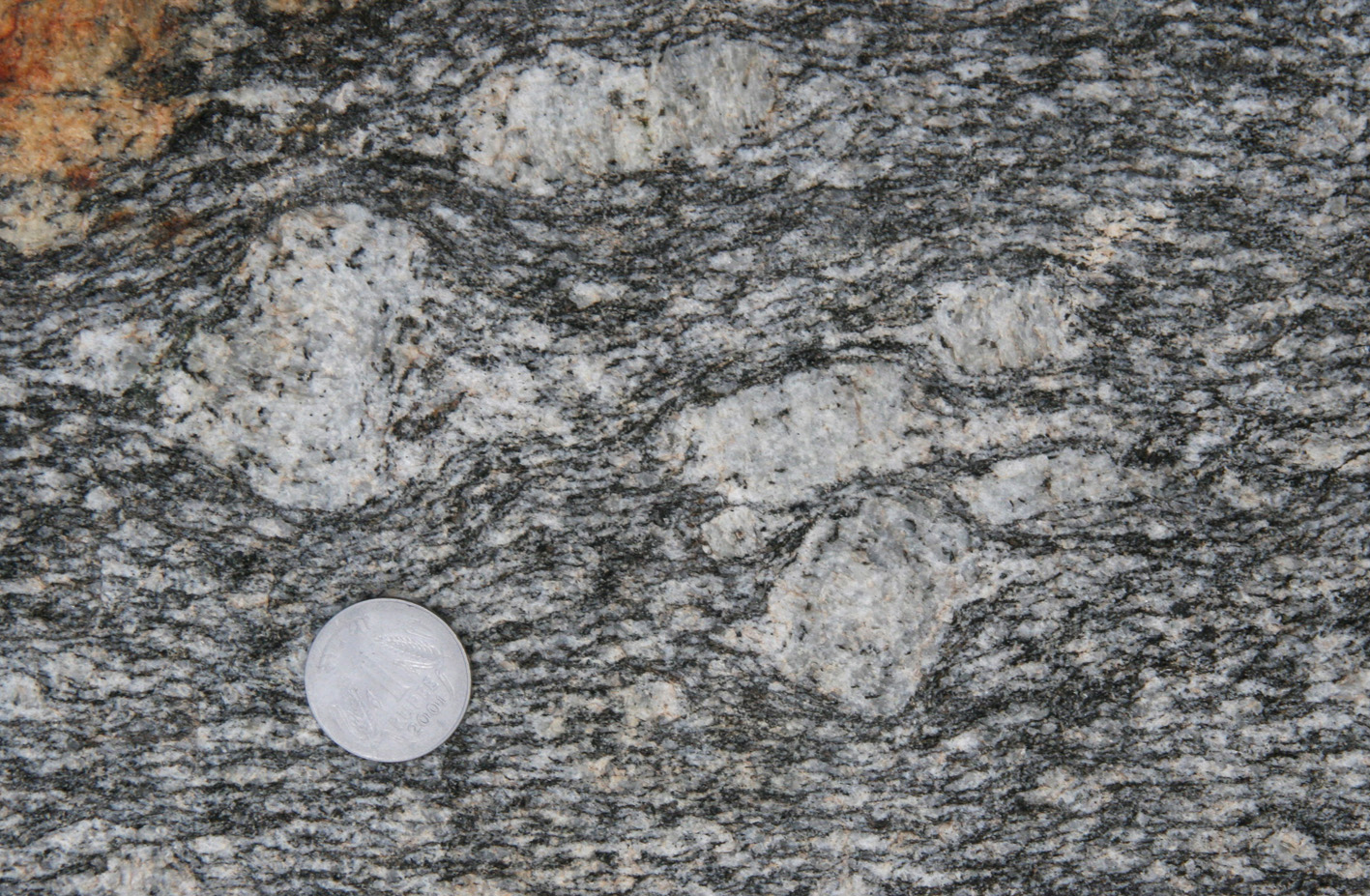
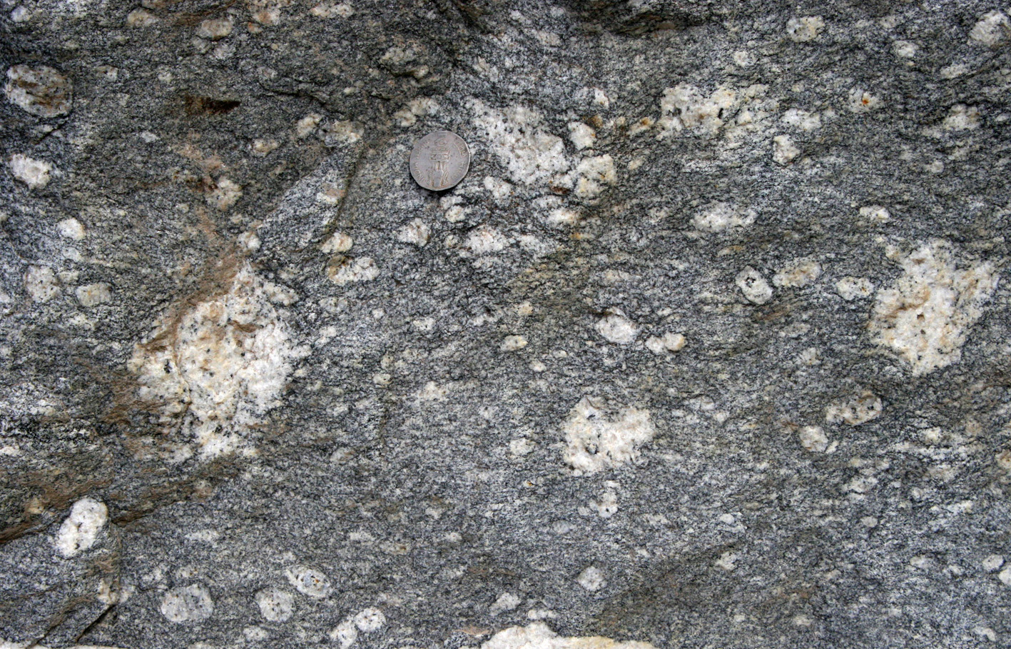
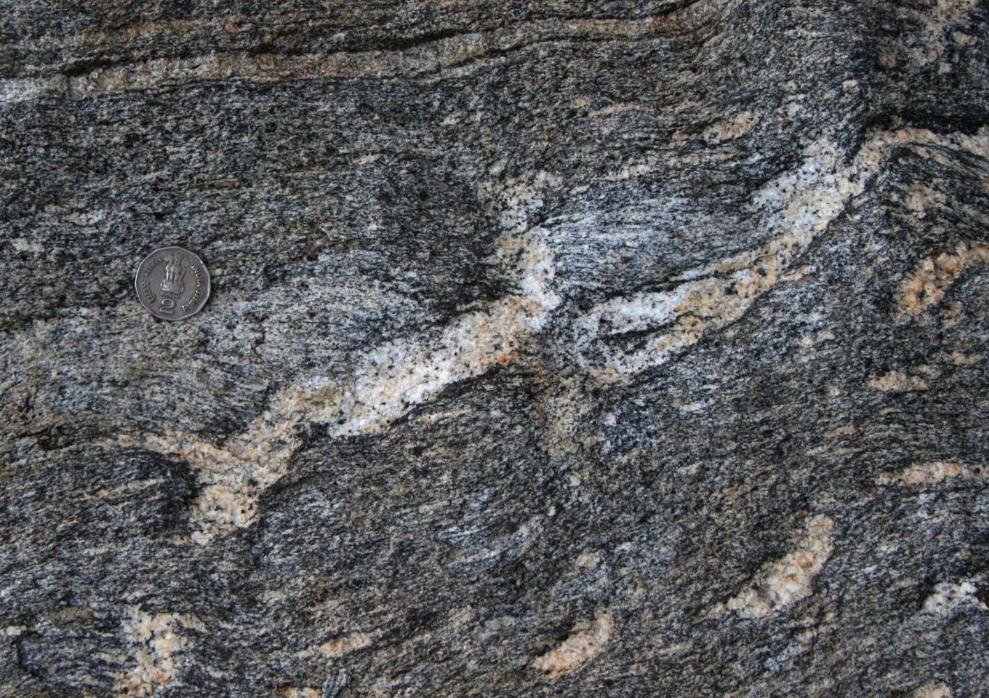
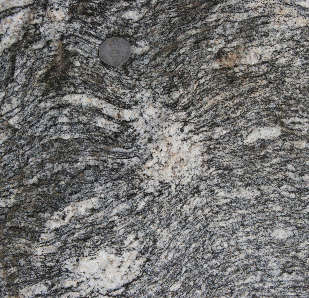
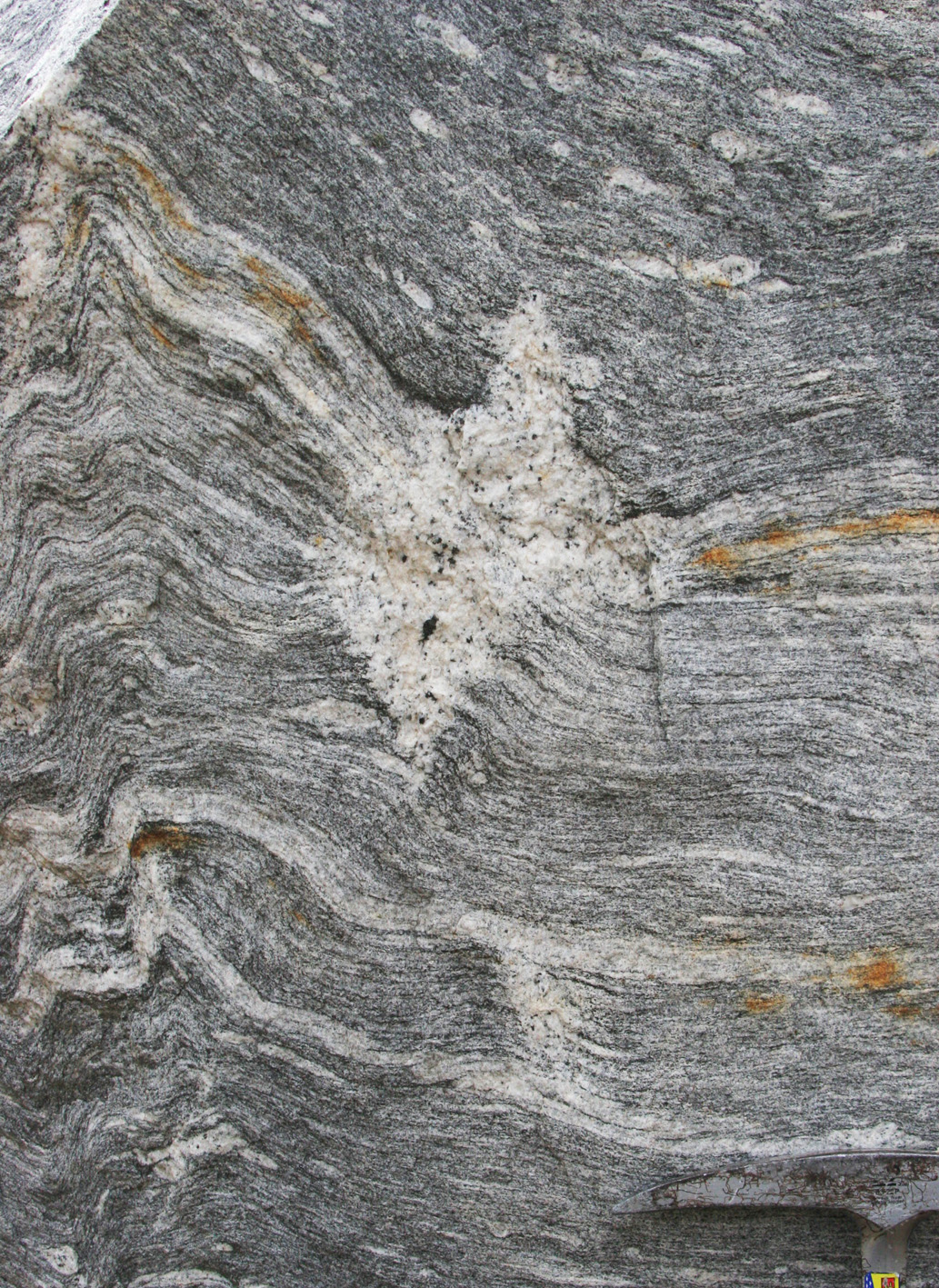
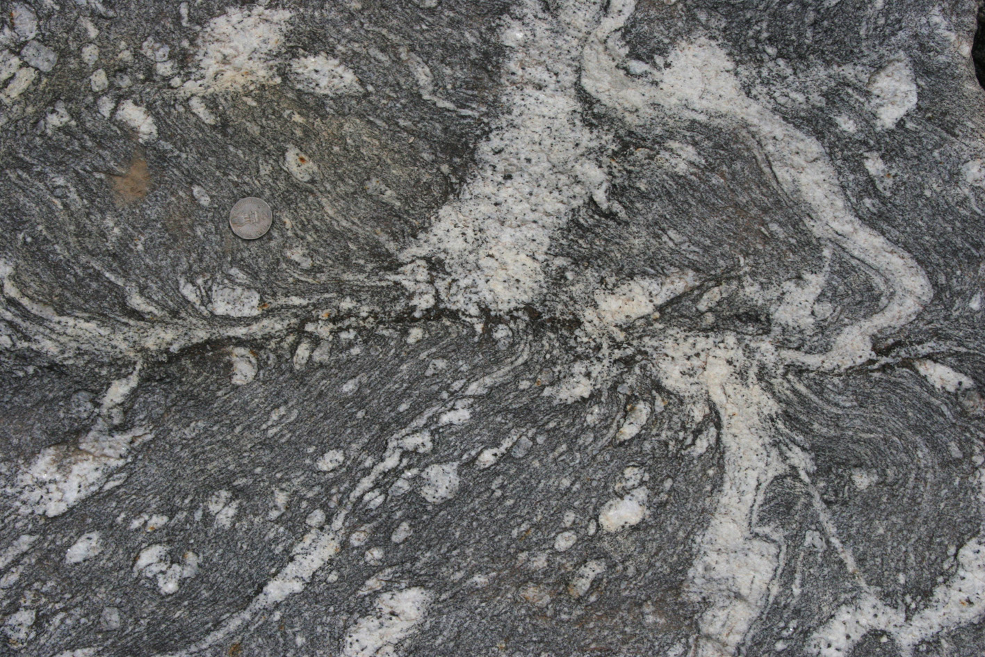
| a) K-feldspar porphyroblasts with asymmetric leucosome tails indicative of dextral shearing. Are the blasts a result of in situ melting or did they pre-date melting? | b) Melt seggregation in a porphyritic gneiss. |
|

|
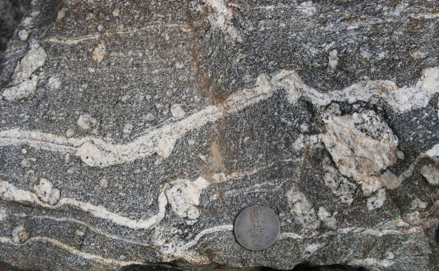
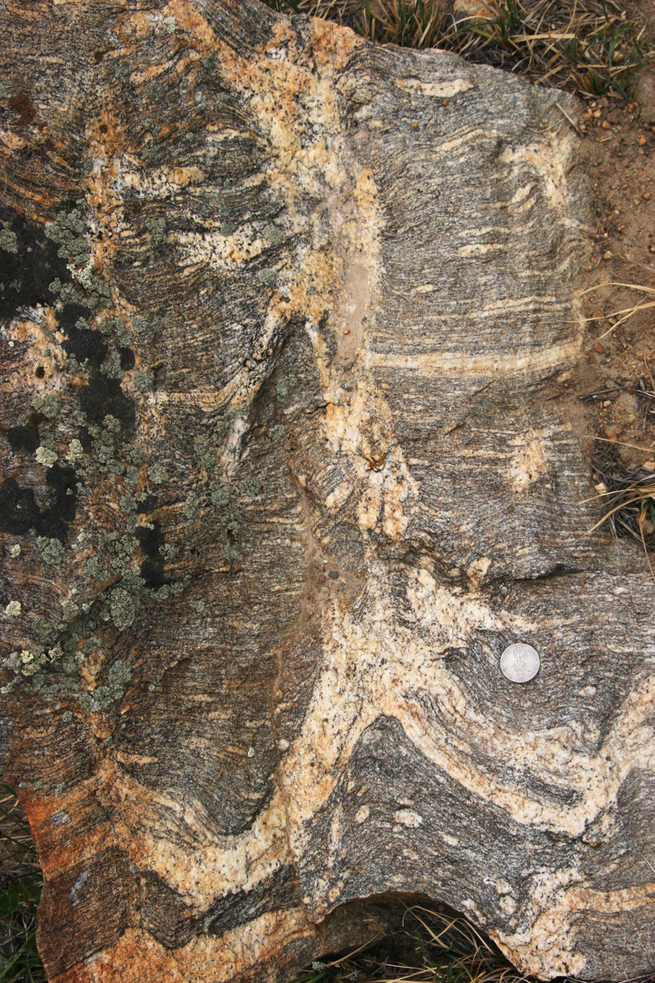
| c) Melt seggregation forming a biotite-speckled leucosome, in a porphyroblastic gneiss. K-feldspar has zoned biotite inclusions. | d) Leucosome network including K-feldspar porphyroblasts. |
|

|
| e) Melt seggregation in a porphyritic gneiss. | f) Melt seggregation in a porphyritic gneiss. |
|
|
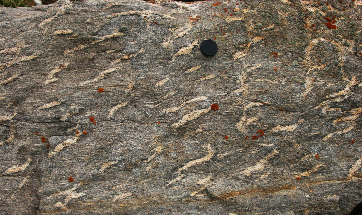
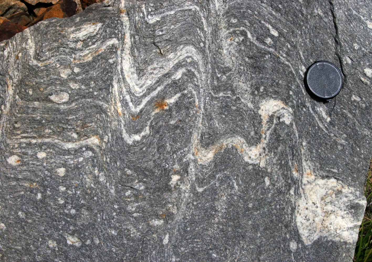
| a) Patchy leucosomes. | b) Patchy leucosomes. An early set of leucosomes is cut by and linked together by a new, less well-delineated (fuzzier) and steeper leucosome. Interpretation: early phase assists with rotation due to shearing, rotation favours the formation of a new set. |
|
|
| c) Patchy leucosomes. An early set of leucosomes is cut by and linked together by a new, less well-delineated (fuzzier) and steeper leucosome. Interpretation: early phase assists with rotation due to shearing, rotation favours the formation of a new set. | d) Patchy leucosomes with typical ends, parallel and perpendicular to foliation. |

|
|
| e) Different styles of leucosomes. There is a central semi-circular area, about 2m across cut by two folded dykes. This region has few small, patchy leucosomes, and is surrounded by a region with many more leucosome patches but where the dykes are missing. This suggests that faint differences in rock type control the volume and behaviour of melt. |
|
|
| a) Patchy leucosome in boudin necks. | b) Patchy leucosome interacting with folds. |

|
|
| a) A certain harmony. Leucosomes and folds in migmatite. | b) Patchy leucosome interacting with folds. |
|
|
| e) Melt accummulation at the contact between two different gneisses. | f) Melt accummulation at the contact between two different gneisses. |

|
|
| a) Antithetic shear zone. This photo shows an antithetic melt-filled, dextral shear zone developed within a broader sinistral shear zone with the shear plane parallel to the horizontal (see asymmetry of clasts). The antithetic shear zone(s) allows for anticlockwise block rotation in response to the broader sinistral shearing. | b) Axial planar foliation acting as shear zones separating blocks with different strain. Some leucosomes are discontinuous across the axial planar foliation. |
|
|
| a) A conjugate pair of melt-filled shear zones, intersecting and allowing for pure shear. |
|
|
| a) Asymmetric folds in leucosome with a narrow melanosome: asymmetry indicates top-to-the-right movement. On the upper part of the same rock, leucosome formed in boudin neck with an axis into the rock, indicative of pure shear and extension parallel to the length of the photograph. The two features cannot result from a single strain axes system and most likely indicate evolving straining during melting. |
|
|
| a) Boudin-like structure interpreted as marking volume decrease due to melt removal. The main axial planar, high strain structure (horizontal) is marked by a melanosome and some leucosomes match and some do not. | b) Fold and an axial planar dyke linked to leucosomes in surrounding foliation. |