Comparisons of Models to Mammalian Teeth
|
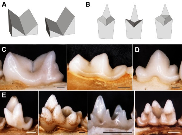
| Comparisons between the models and real mammalian tooth forms. L-R. Model tools: A, single-bladed tools, symmetrical (2i) and asymmetrical (2ii); B, double-bladed tools (3i, 3ii, 4ii). Mammalian tooth forms: C, lower carnassials of Felis catus (Carnivora: Felidae) and Mustela frenata (Carnivora: Mustelidae); C, premolar of F. catus; E, lower molars of Tenrec ecaudatus (Insectivora: Tenrecidae), Didelphis virginiana (Didelphimorphia: Didelphidae) and Chalinolobus gouldii (Chiroptera: Vespertilionidae), and upper molar of Desmana moschata (Insectivora: Talpidae). Scale bar is 1 mm. |
|
|
Alistair Evans,
May 2003