Contents:
b) Water-fluxed melting: no peritectic minerals
c) Bt-dehydration melting
d) Bt-dehydration leucosomes overprinted by Tml-Ms-Bt-Gnt (water-fluxed) leucosomes
e) Early Bt-leucomes related to water-fluxed anatexis overprinted by Tml-Ms-Bt-Gnt (water-fluxed) leucosomes
f) Migration of Tml-Ms-Bt-Gnt leucogranite
a) Major rock types in the valley
b) Water-fluxed melting: no peritectic minerals
c) Bt-dehydration melting
d) Bt-dehydration leucosomes overprinted by Tml-Ms-Bt-Gnt (water-fluxed) leucosomes
e) Early Bt-leucomes related to water-fluxed anatexis overprinted by Tml-Ms-Bt-Gnt (water-fluxed) leucosomes
f) Migration of Tml-Ms-Bt-Gnt leucogranite
Reru valley shows rocks with two phases of melting. In parts of the
valley the first phase of anatexis is associated with biotite
dehydration melting. In other parts, with water-fluxing melting giving
rise to a Bt-leucosome in granitic rocks lacking obvious peritectic
minerals. The two are mutually exclusive. This first phase of
water-fluxed anatexis is syn-kinematic.
These two events are followed by a widespread second anatectic phase
characterized by B-rich water influx producing Tml-Ms-Bt-Gt
leucogranites. This is syn- to post-kynematic.
b) Water-fluxed melting: no peritectic minerals such as garnet or
sillimanite, passive biotite, voluminous leucosomes. Two main types:
an early event characterized by Bt-bearing leucosomes (figures 3a-c), a
later event characterized by Tml-Ms-Gnt-Bt leucosomes (figures 3d-f).
In this subsection, only one anatectic event is obvious. In section d)
and e) Iexplore overpriting relationships.
c) Bt-dehydration melting
d) Bt-dehydration leucosomes overprinted by Tml-Ms-Bt-Gnt
(water-fluxed) leucosomes
e) Early Bt-leucomes related to water-fluxed anatexis overprinted by
Tml-Ms-Bt-Gnt (water-fluxed) leucosomes
f) Migration of Tml-Ms-Bt-Gnt leucogranite: this section expands on
some of the syn-kinematic extraction features already described above
such as concentration
in boudin necks, shear planes associated with folds, and axial planar
segregations
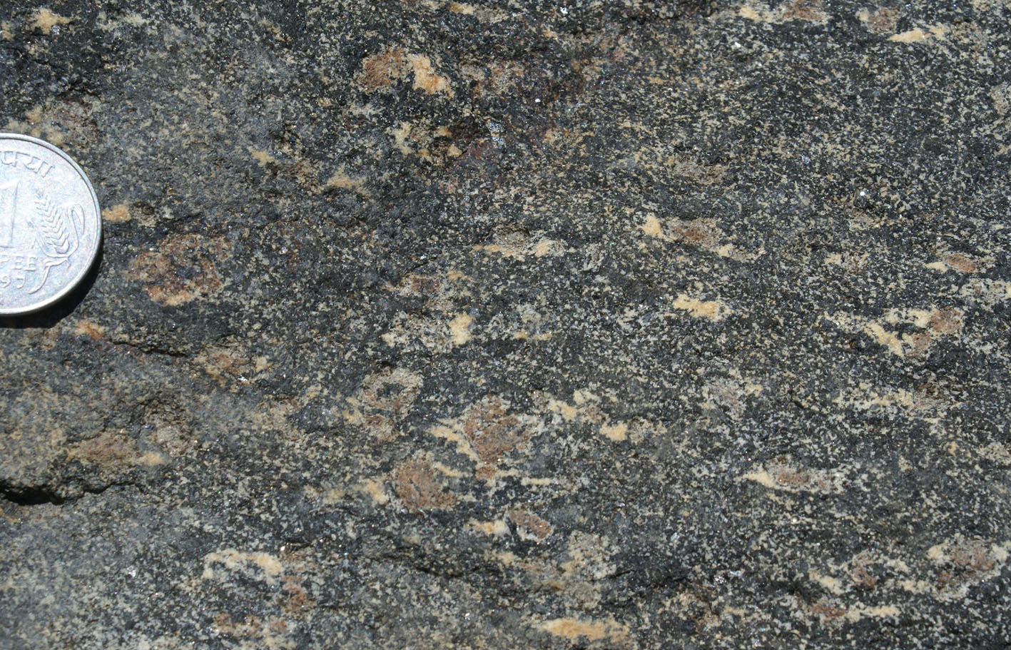
a) Major rock types in the valley Phase 1 dehydration followed by Phase 2 B-rich water-fluxed melting
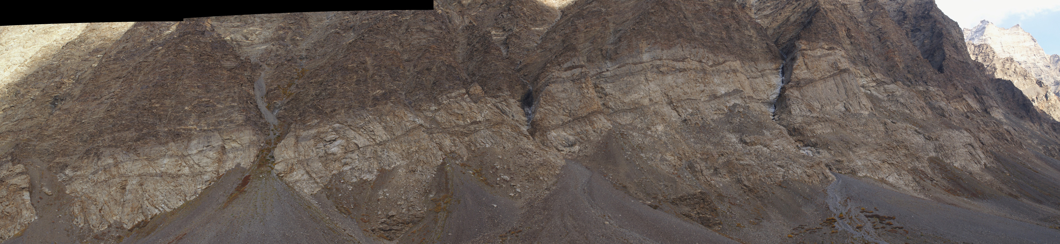 Figure 1. East side of the Reru valley. A thick and continuous layer-parallel leucogranite sill >9 km long (field of view 6km horizontal length) embedded in migmatites with numerous sills and dykes. Immediatelly above the sill,sills and dykes are relatively rare and increase in number away from the large sill further up the wall. This suggests that the sill may have provided a sink for surrounding magma. |

|
|
|
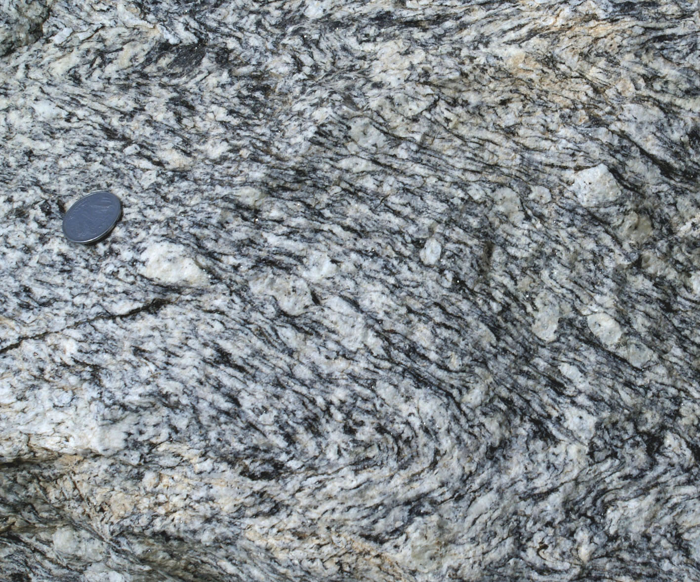
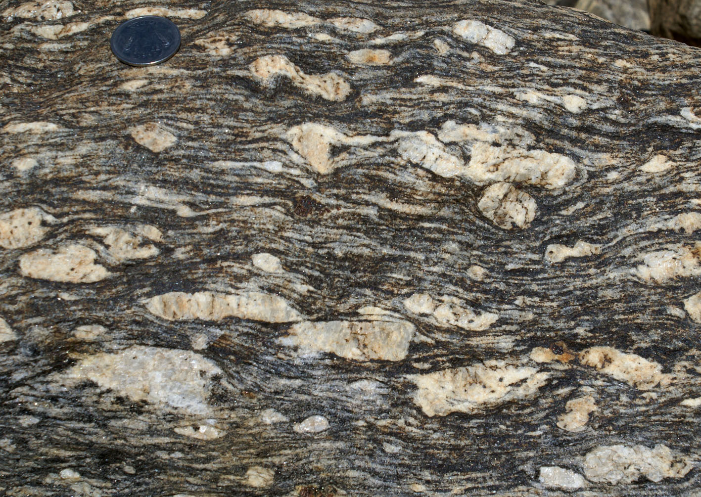
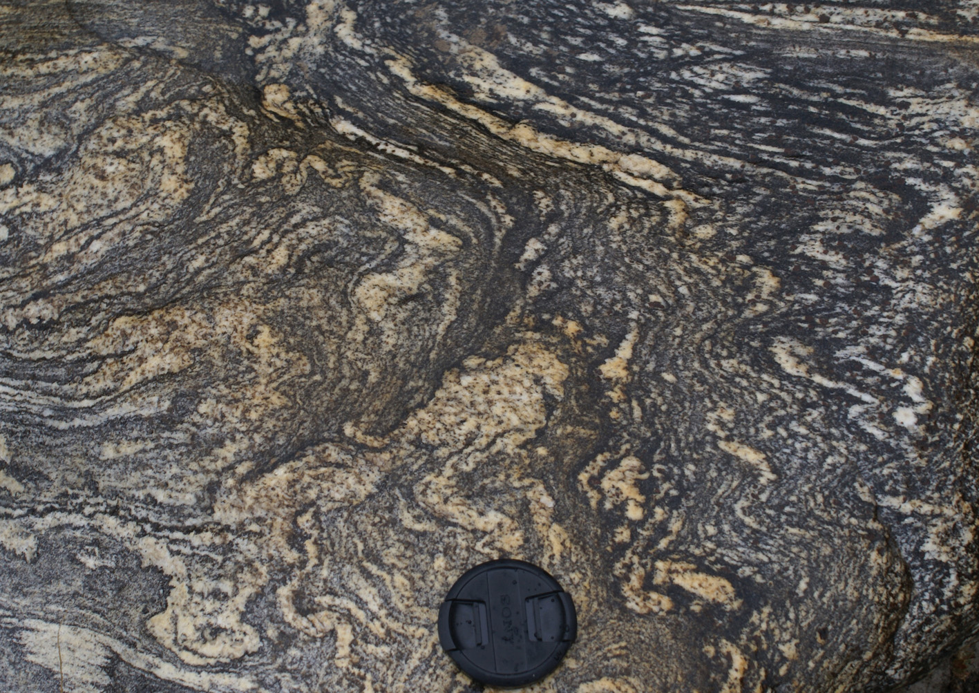
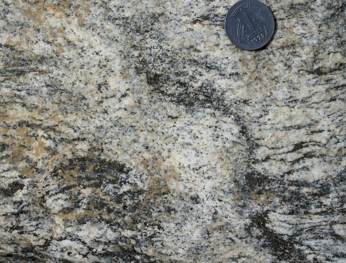
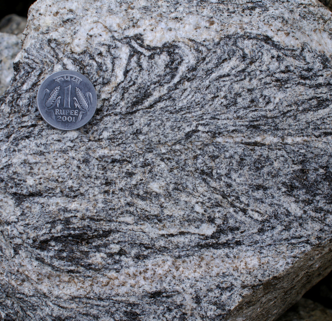
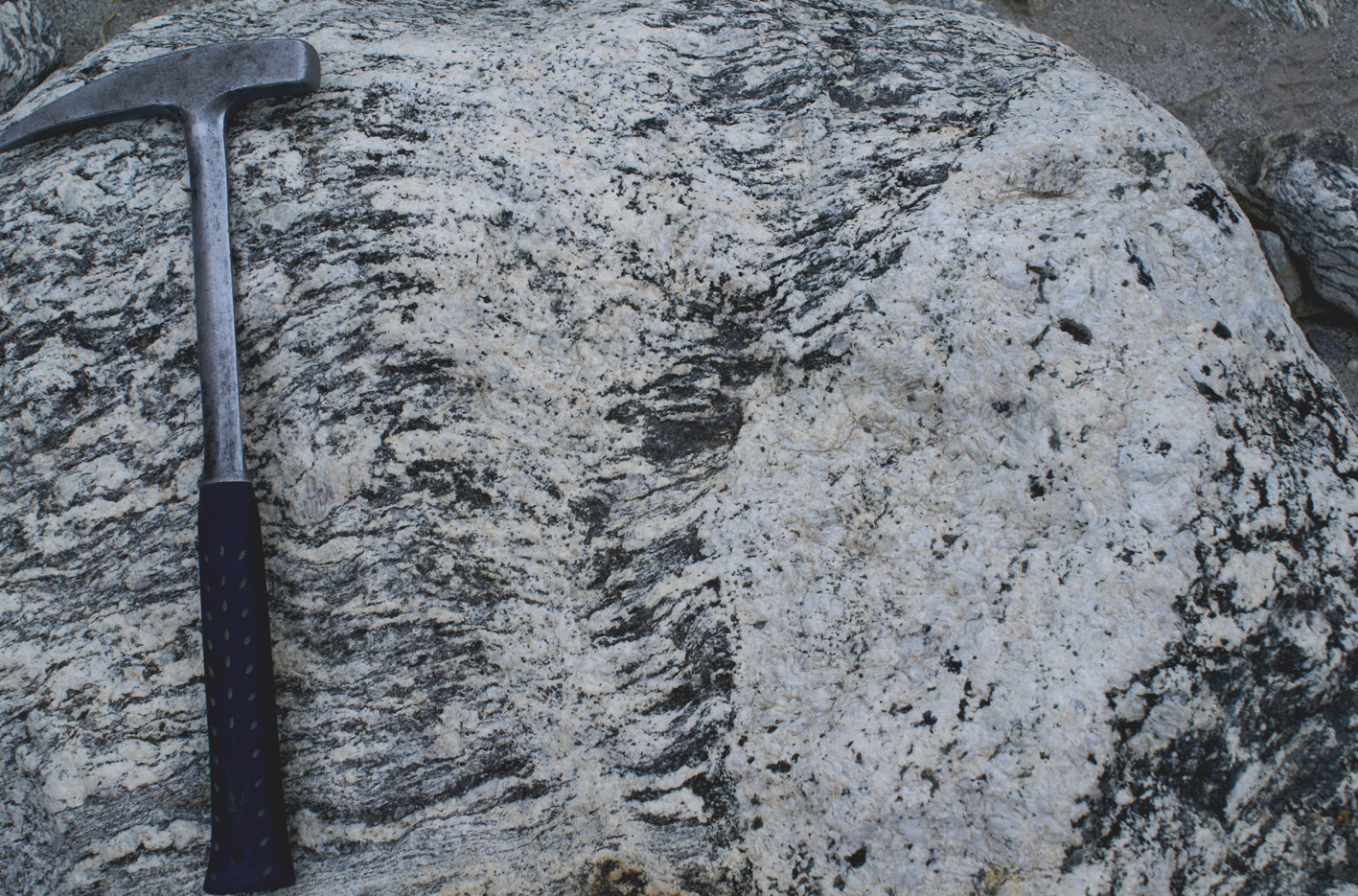
| Figure 2a) Two sides of typical Ms-Bt mylonitic granite showing intense lineation. | Figure 2b) Folded coarse gneiss | Figure 2c) Coarse-grained mylonite with K-feldspar porphyroblasts whose protolith was a porphyritic granite . |
 |
 |
|
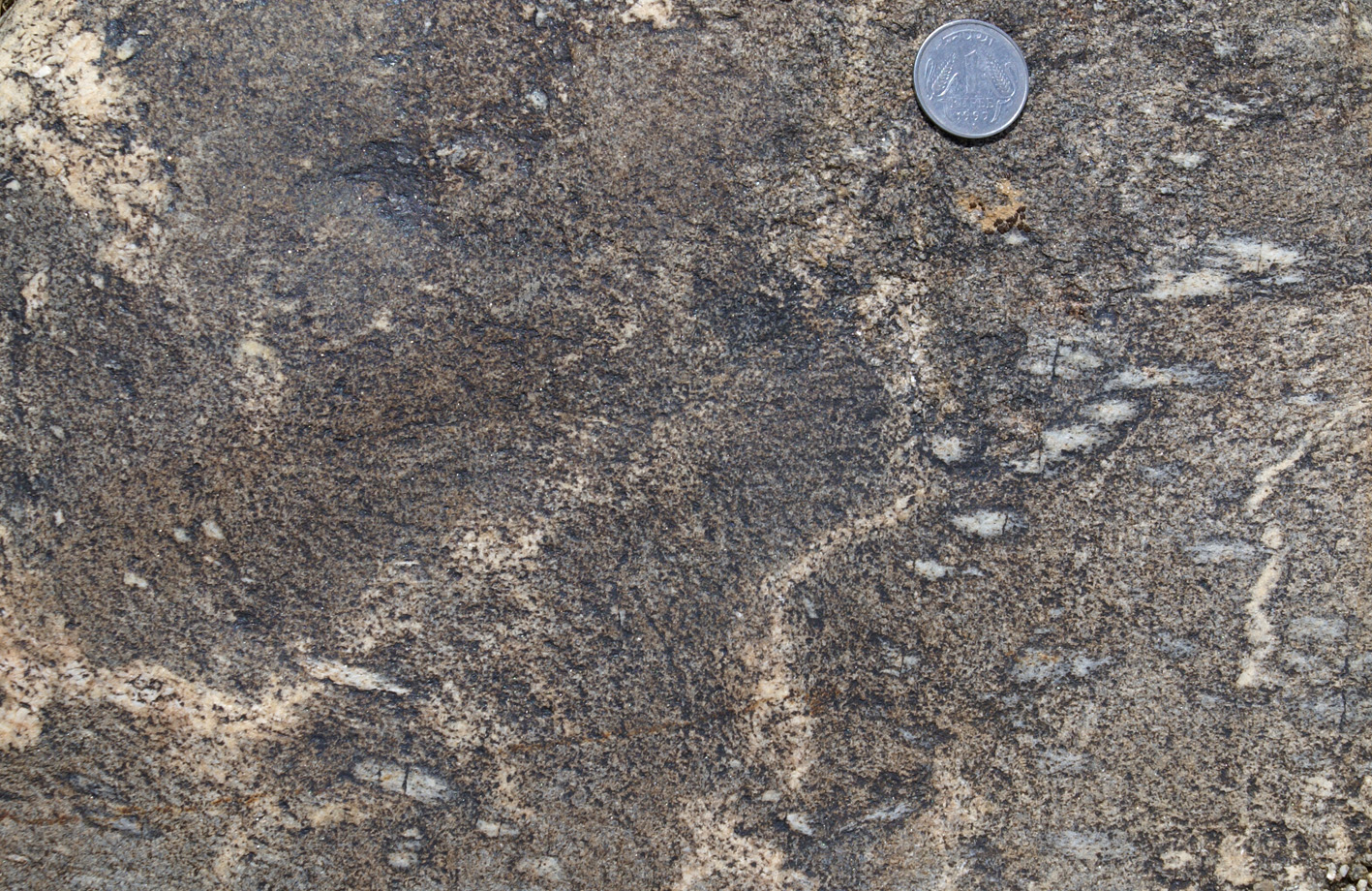
| Figure 2d) Sillimanite-biotite schist with leucosome having diffuse boundaries suggesting in situ origin. Lack of spatial relationship with sillimanite suggest that melting is unrelated to the presence of sillimanite. | Figure 2e) Folded sillimanite-biotite schist intruded by tourmaline leucogranite with irregular boundaries and narrow melanosome rim in places. Rock face close to parallel with foliation. The leucogranite is unrelated to sillimanite grains (left of coin). | Figure 2f) Folded biotite-rich diatexite with refractory layers. |

|
|
|
| Figure 2g) Tml-leucogranite diatexite. | Figure 2h) Tml-Ms pegmatite resulting from melting. | Figure 2i) Banded tourmaline leucogranite. |
 |
 |
 |
| Figure 3a) Leucosome surrounded by a narrow melanosome rim indicative of in situ derivation, in biotite granite. The leucosome is ptygmatically folded and streteched in the third dimension (below and to the left of the coin in this curved block) suggesting a constrictional environment at high temperature (possibly during melting). | Figure 3b) Migmatite with ~40% leucosomes, lacking anhydrous peritectic minerals, and strongly folded and stretched in the thrid dimension on the top part of the photo (curved block). | Figure 3c) Leucosome linked to layer parallel leucosome and dragging refractory biotite grains that accumulate irregularly at the side of the cross cuttting leucosome. |
 |
 |
 |
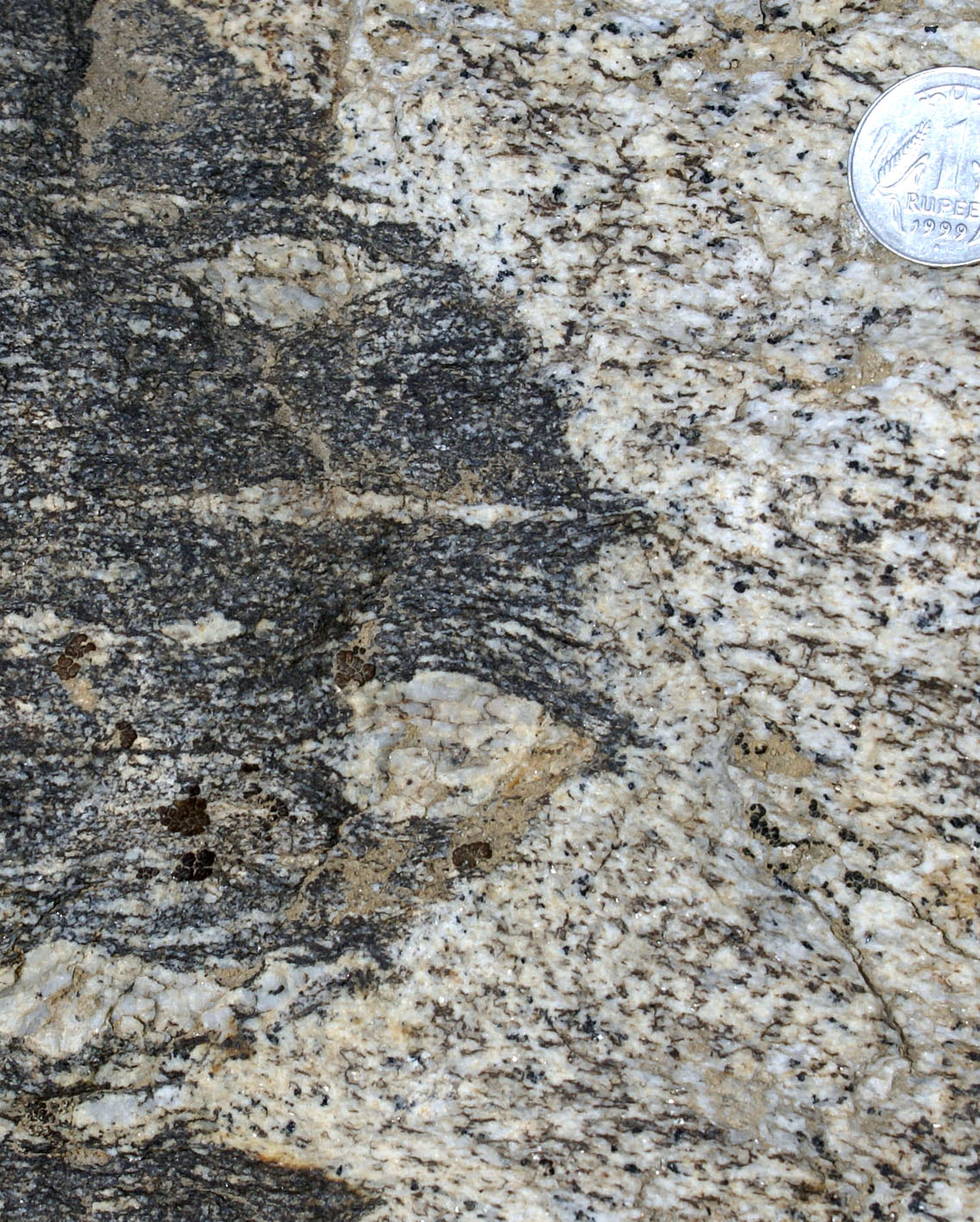
| Figure 3d) Axial planar muscovite-bearing leucosomes linked with leucosomes in the intervening folds, indicative of syn-kinematic melting. | Figure 3e) Two cross-cutting tourmaline-bearing leucosomes in granitic migmatite. The one on the left shows intricate relationships with smaller, foliation parallel leucoscomes in the surrounding rock suggesting mainly in situ generation, whereas the one on the right has planar margins with local interaction with surrounding leucosomes, suggesting mainly intrusive nature. The large volume of leucosomes and the lack of sillimanite, garnet or any other obvious peritectic mineral suggests water-fluxed melting. | Figure 3f) Tortuous white vein with black tourmaline in Bt-leucogranite with a pre-existing foliation. In this same rock there are equigranular, centimetric patches of unfoliated tml-leucogranite suggesting in situ derivation. |
 |
 |
 |
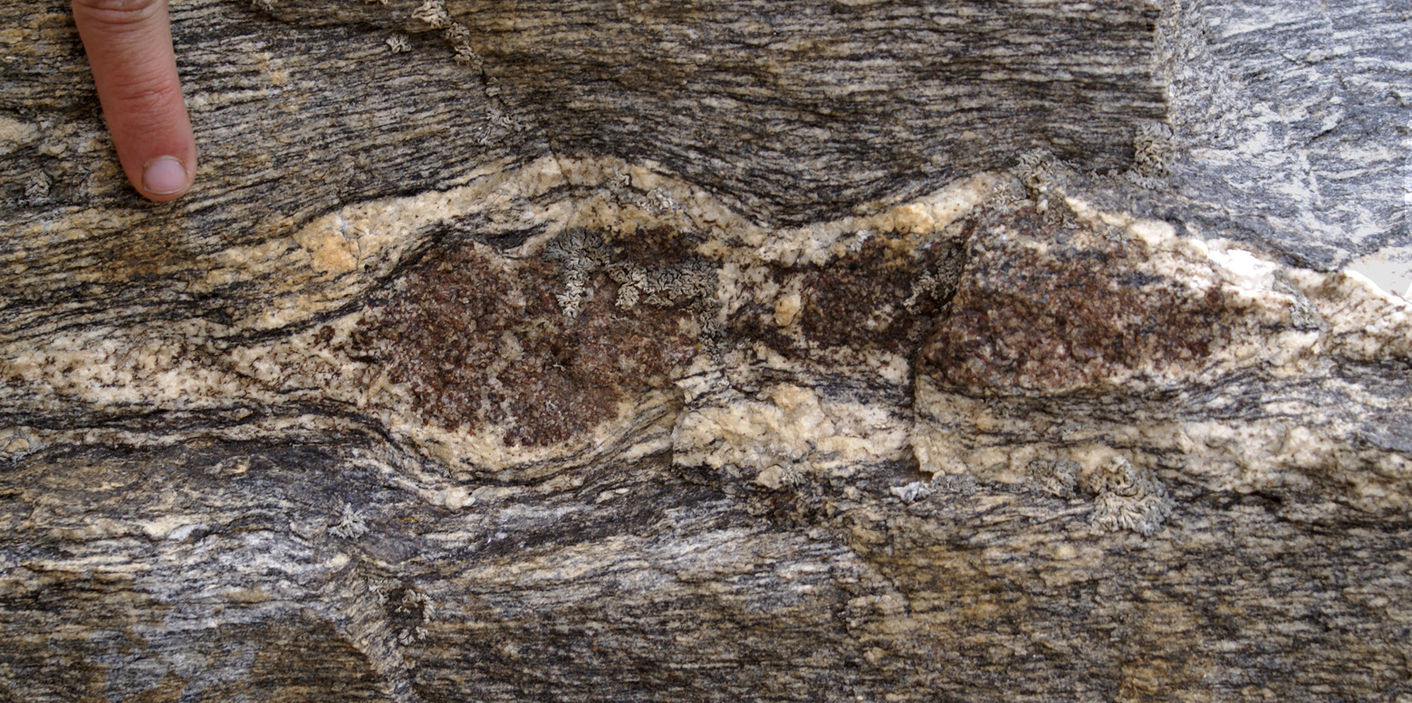
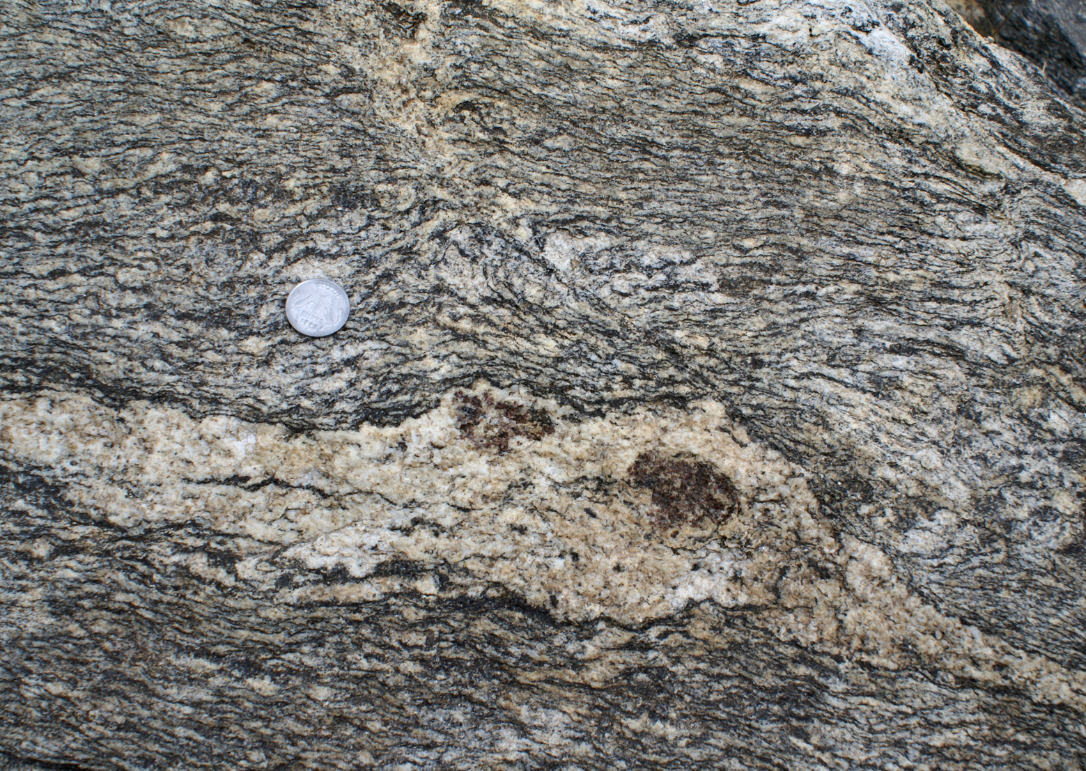
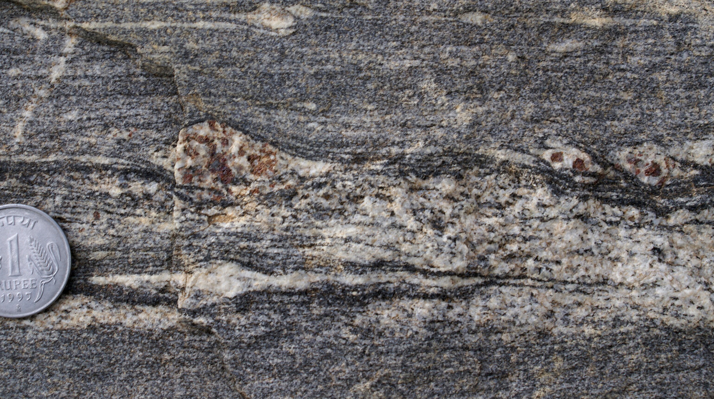
| Figure 4a) Leucosome including large garnets in Bt-granite. Plane in rock nearly parallel to foliation, enhancing the size of features. | Figure 4b) Large garnet grains surrounded by leucosomes. The relatively small volume of leucosome compared to the garnet, and the absence of garnet retrogression to biotite, suggest loss of melt from the system. | Figure 4c) Small garnets surrounded by narrow leucosomes, remnants of magma. |
 |
 |
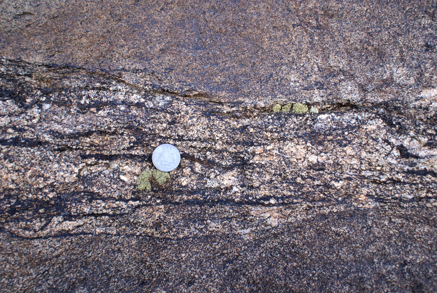
| Figure 5a) Garnet in leucosome parallel to the main foliation localizing strain during a late (or continued) deformation event. | Figure 5b) Garnet in leucosome forming apaprent sinistral fish indicative of pre- or syn-kinematic partial melting through Bt-dehydration. |
 |
 |
 |
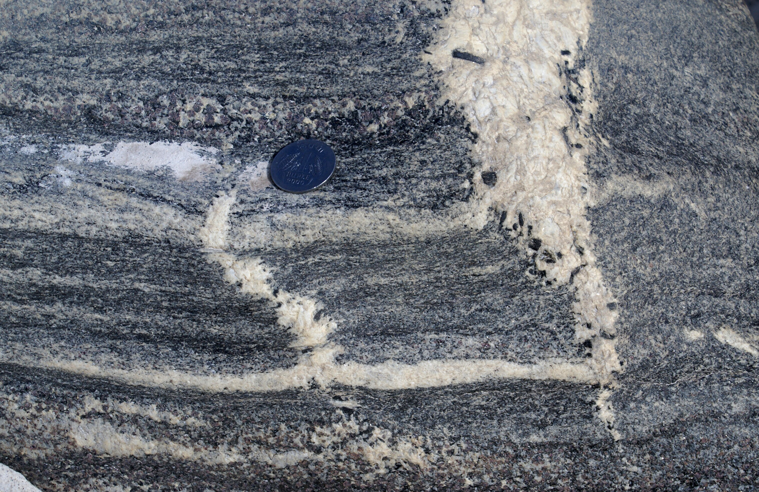
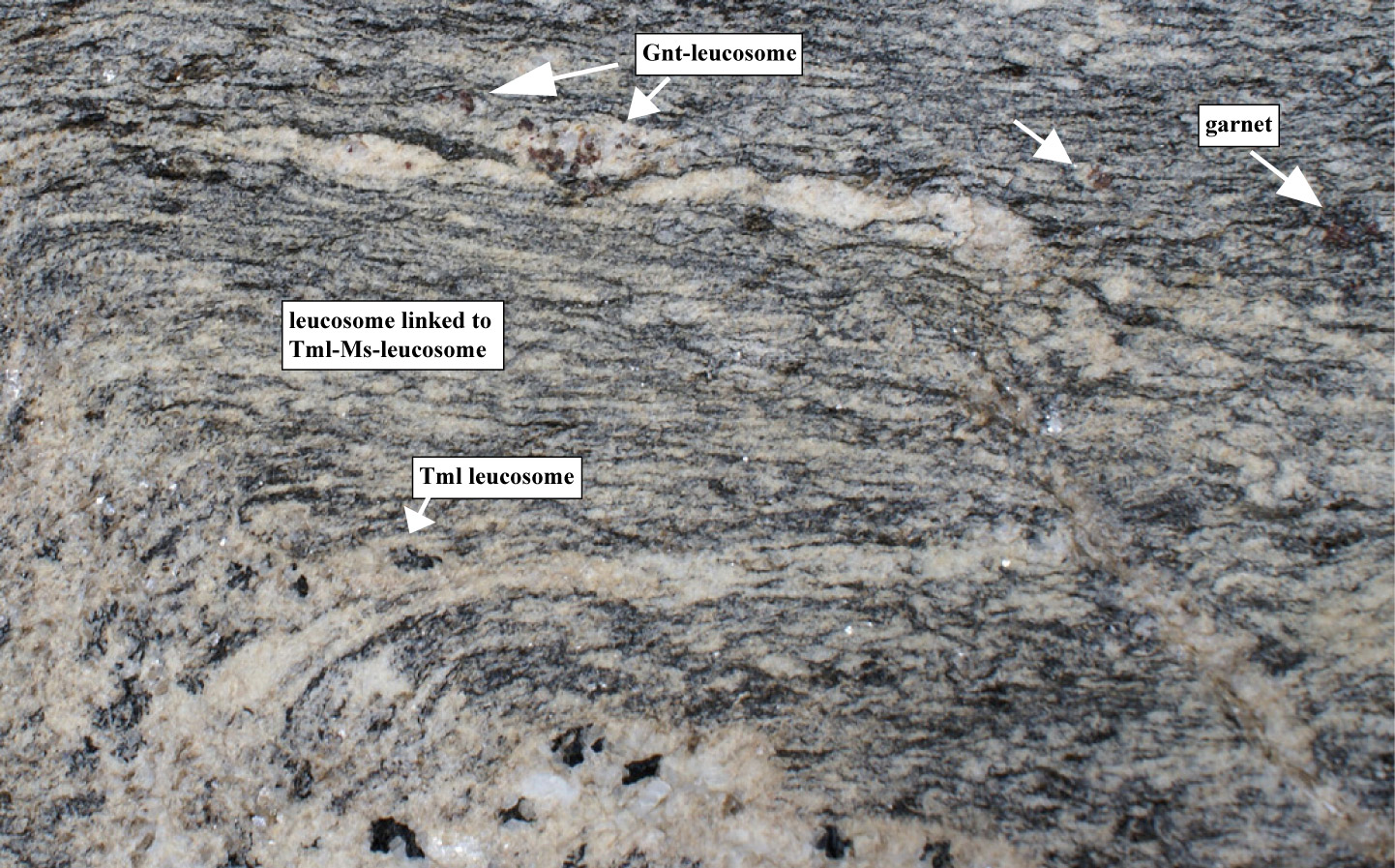
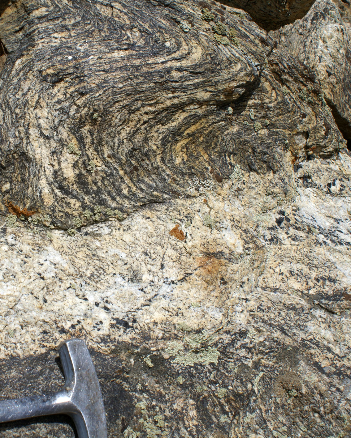
| Figure 6a) Garnet and its leucosome crenulated in hinge zone of late folding . Tml-rich leucosomes are axial planar to this fold and fills in boudins in limbs (Figures 6c). | Figure 6b) Tml-leucogranite leucosome with diffuse boundaries with surroundings (in situ) but also cross-cutting strong foliation and layering, including layers rich in garnet-sillimanite-biotite and associated leucosomes, suggesting an earlier partial melting event. | Figure 6c) Same outcrop as Fig. 6a, showing a leucosome in boudin neck with large tourmaline grain linked to Ms-bearing leucosome system (parallel and left of finger, and then parallel to foliation. This represents melt segregation in the limb of the fold that crenulated early formed, garnet-bearing leucosomes (Fig. 6a). |

|
|
|
| Figure 6d and e) Segregation of Ms-Tml-Bt leucosomes into a thrust plane that lubricates a fold-thrust. Melt in the thrust plane is linked continuously with melt in folde leucosomes suggesting in situ derivation during deformation. This melting event is mineralogically and temporally distinct to the one producing the garnet-leucosome association depicted in Fig. 6a. d) Depitcs an early stage of development of a melt-lubricated fold-thrust, whereas e) depicts a late stage characterized by tighter fold and better defined leucosome seggregation. Same outcrop as Fig. 6a and c. | Figure 6e) Same as d but a later stage of evolution of a melt-lubricated fold-thrust. | Figure 6f) Migmatitic gneiss with coarse-grained felsic leucosome lenses including minor Bt, cut across by a finer-grained Tml-Bt granite. This granite has interfingering contacts with the migmatite suggesting local derivation, at least in part. Also the granite has the same E-W foliation as the gneiss, suggesting that it is contemporaneous with or older than the deformation event that produced such foliation. |
 |
 |
 |
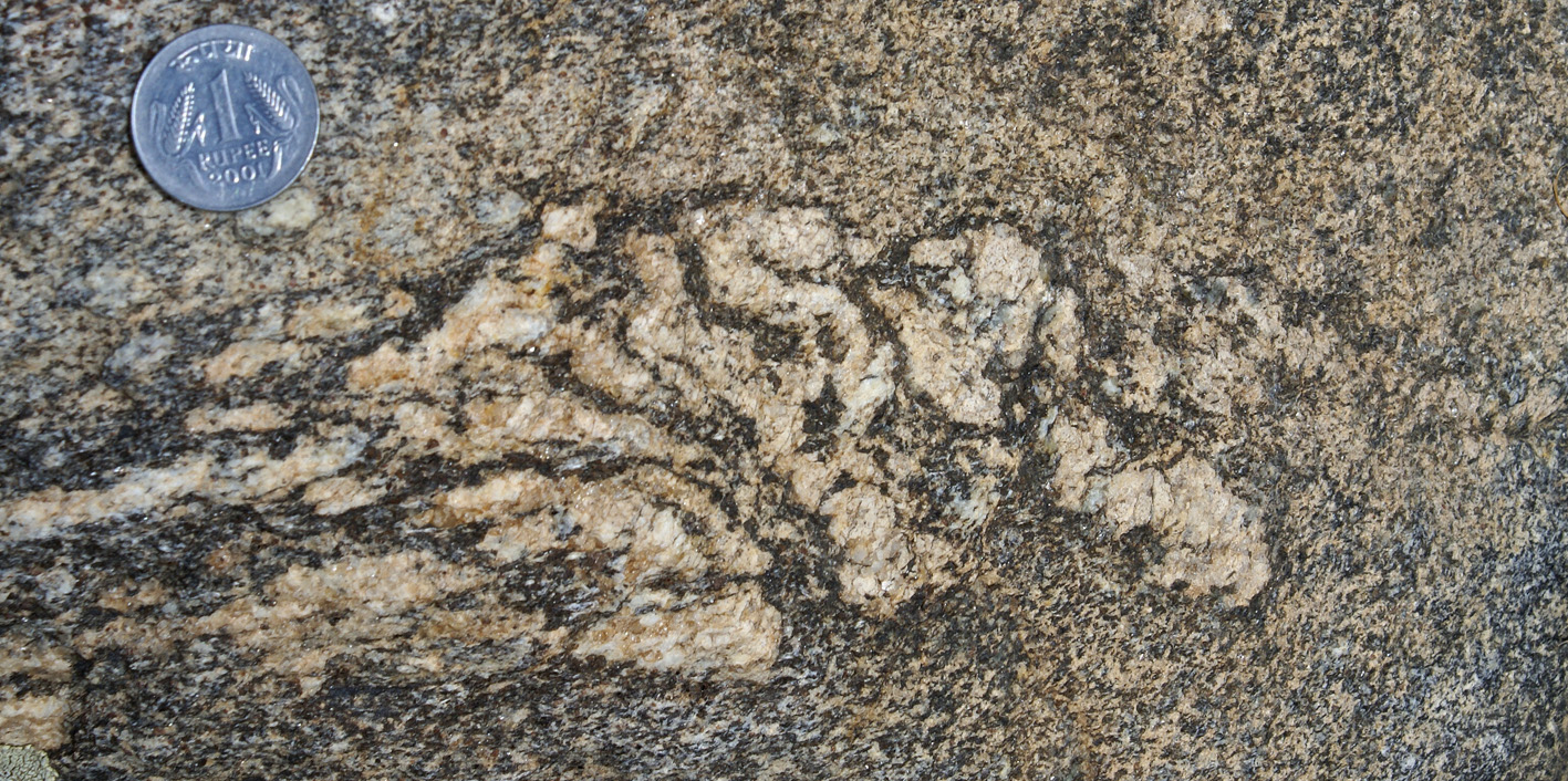
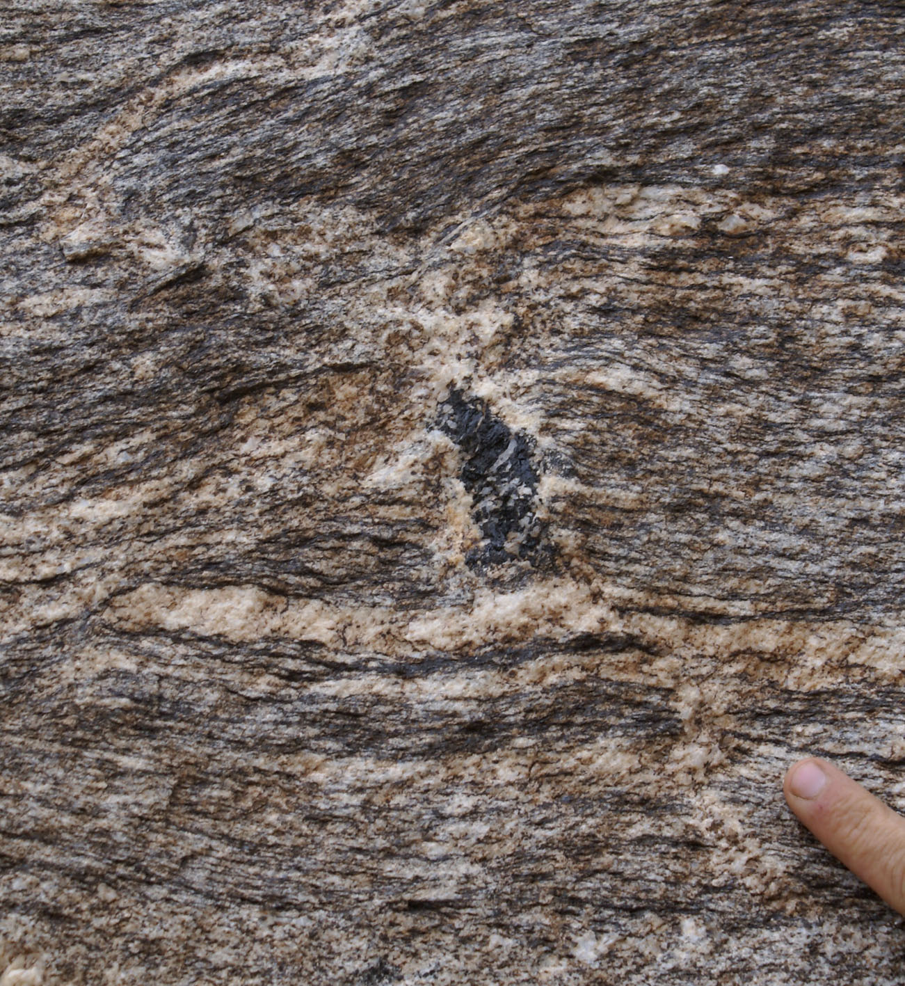
| Figure 7a) Segregation of Tml-Ms leucogranite into an incipient fold hing, from foliation-parallel leucosomes in the granite. Granite has garnet in small leucosomes not physically linked to the network that feeds the leucogranite segregation. These are interpreted to represent an early anatectic phase of Bt-dehydration melting. | Figure 7b) Detail of a. | Figure 7c) Similarly to Figure 7a, Tml-Bt-Ms leucogranite dyke linked continuously a foliation-parallel leucosome lacking garnet, in a rock with numerous garnets in leucosomes interpreted to pre-date the dyke. |
 |
 |
 |
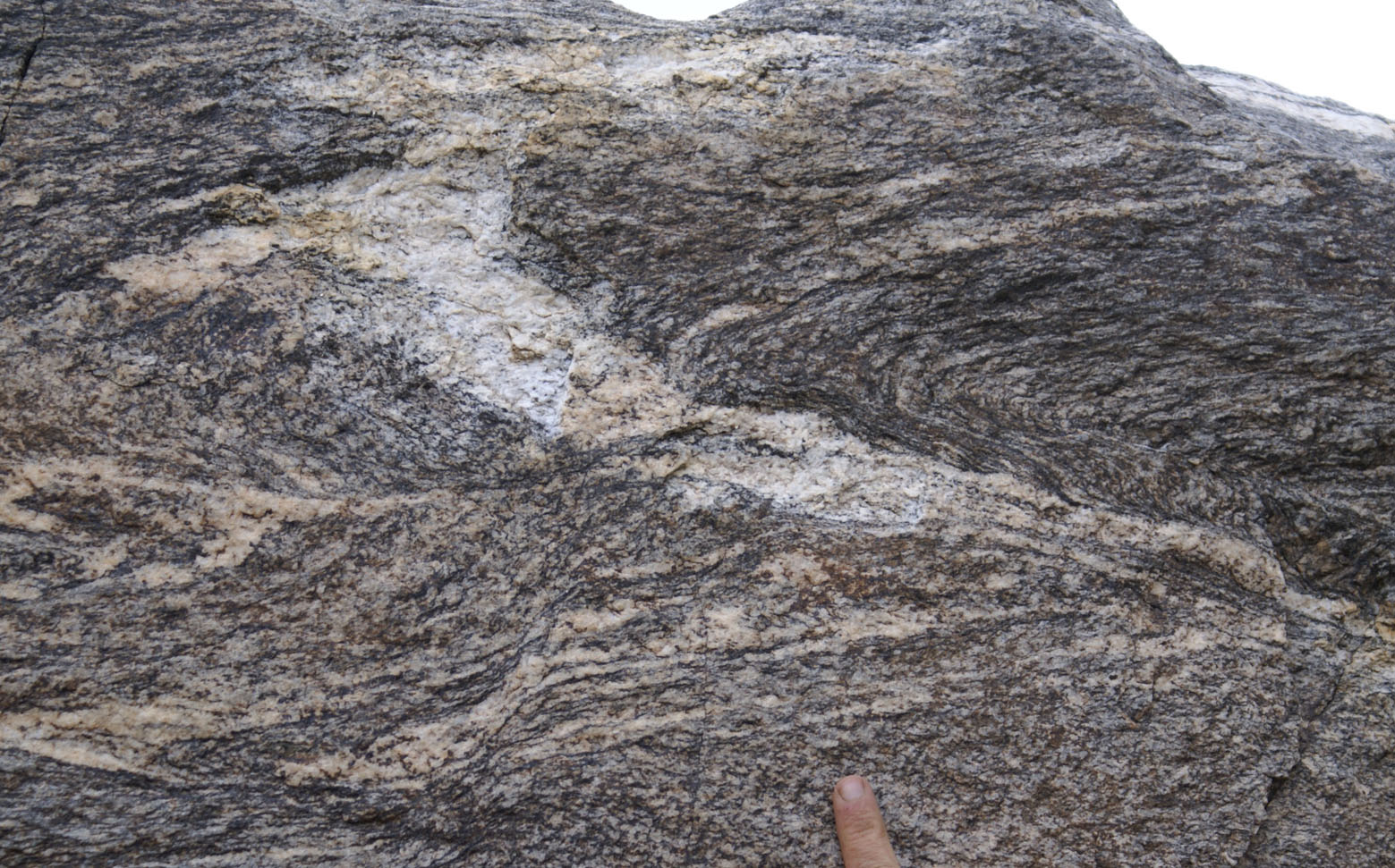
| Figure 8a) Ms-Bt migmatitic gneiss with lens-shaped leucosomes parallel to a folded foliation (interpreted to represent the first anatectic event lacking Tml or peritectic minerals). This is cross-cut by a later phase of Tml-Bt leucogranite which is linked continuously with a Bt-leucogranite dykes (horizontal band along the bottom and top of the photograph). This late phase has diffuse boundaries against the surrounding migmatite in one part (black arrow), and a cross-cutting relationship boundary in another (dashed arrow). This relationship can be interpreted either as a late tectonic Tml-Bt bearing melting event overprinting a pre-existing, but still unconsolidated early anatectic event, or a pre-existing, re-solidified migmatite. | Figure 8b) Folded migmatite cut across by late Tml-leucogranite (~axial planar to folds). | Figure 8c) . |
 |
 |
 |
| Figure 9a) Layered dyke characterized by melanocratic bands and leucocratic bands interpreted to represent mobilization of a diatexite. This dyke was linked in the field with a patch migmatite, such as the one in the upper right corner of the photograph. Lack of peritectic minerals or tourmaline or muscovite suggest that this represents the first water-fluxed anatectic event characterized by accumulation of refractory Bt and Qtz in melanosomes. | Figure 9b) Detail of a). | Figure 9c) In the same block as a) and b) there are local patch leucosome with large tourmaline grains. The relative timing in relation to the biotite is undefined in this block. |
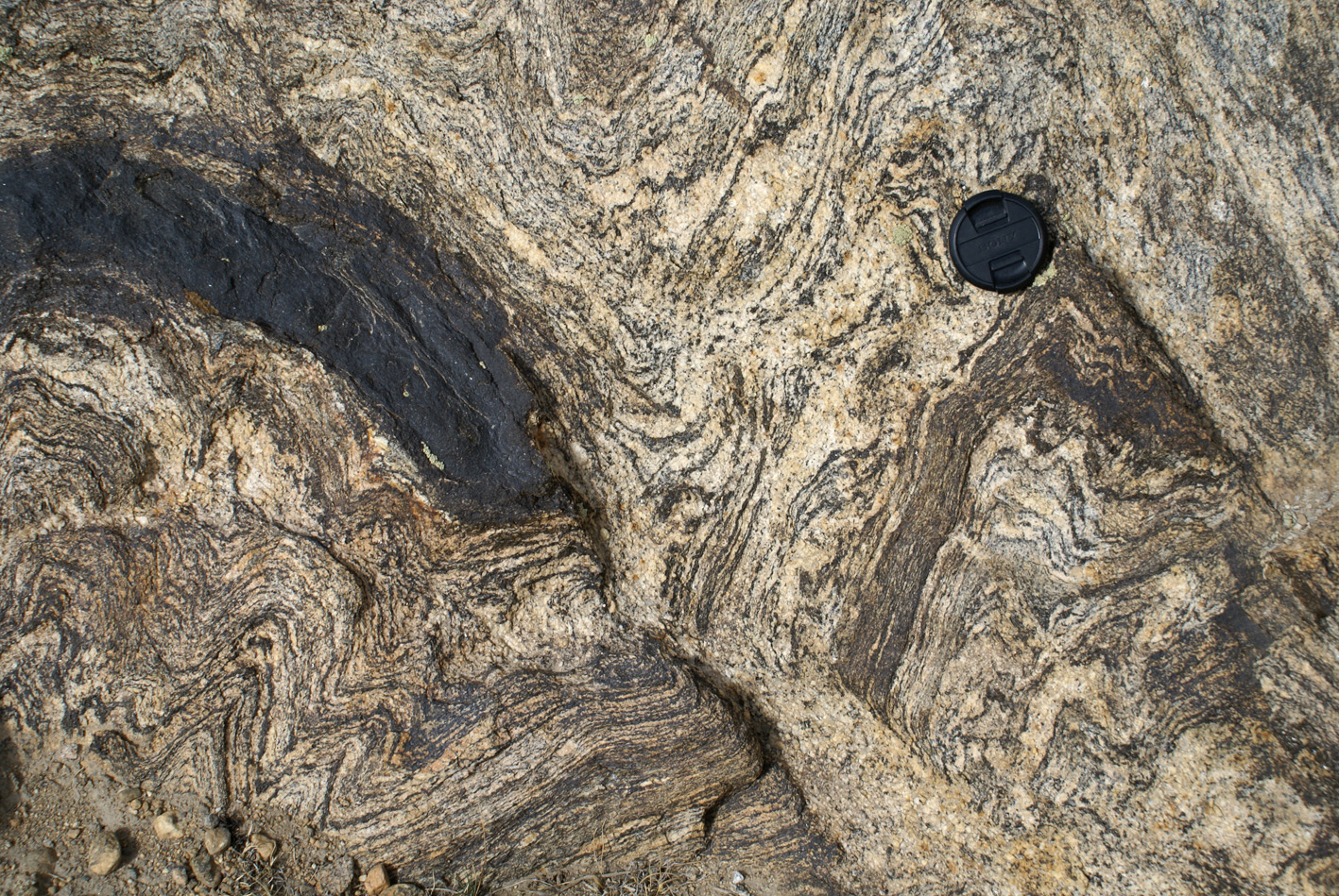
 Figure 10) Extraction of Tml-leucosomes through limbs and axial planes from a folded granitic gneiss. |