Peritectic Hornblende: Water-Assisted Melting of Biotite-Amphibolite, Ladakh, NW India
|
Muglib
Tng200 show here:
a) pattern of melting, 4763, 4767, 4780, 4751 and detail in 4755 , 875, 933, 935, 915, 4770
b) break up of amphibolite to form diatexite stitch 925-926, show also 924, DSC00921good
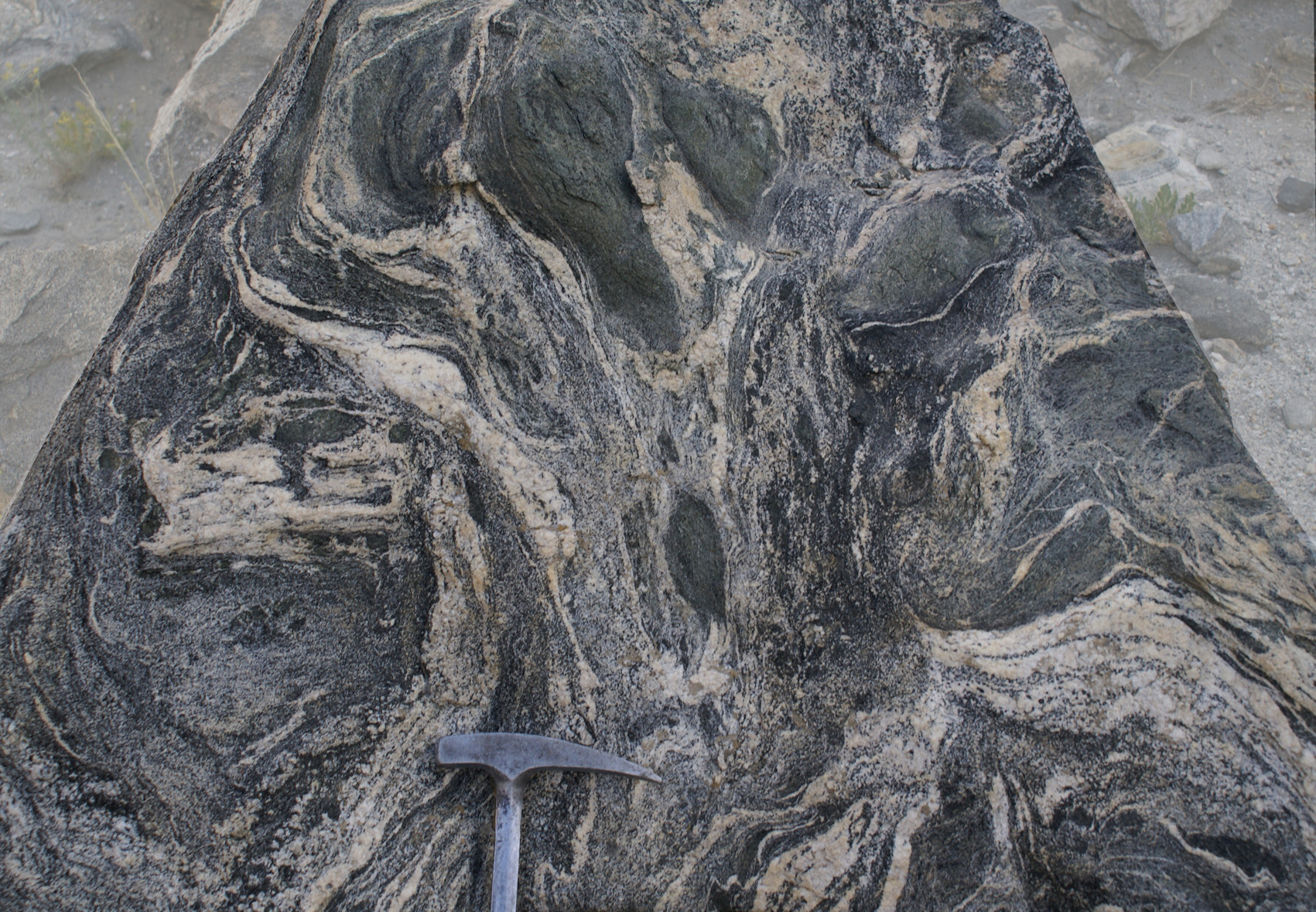
c) folding and melting 885good, DSC00888zoom, 1216
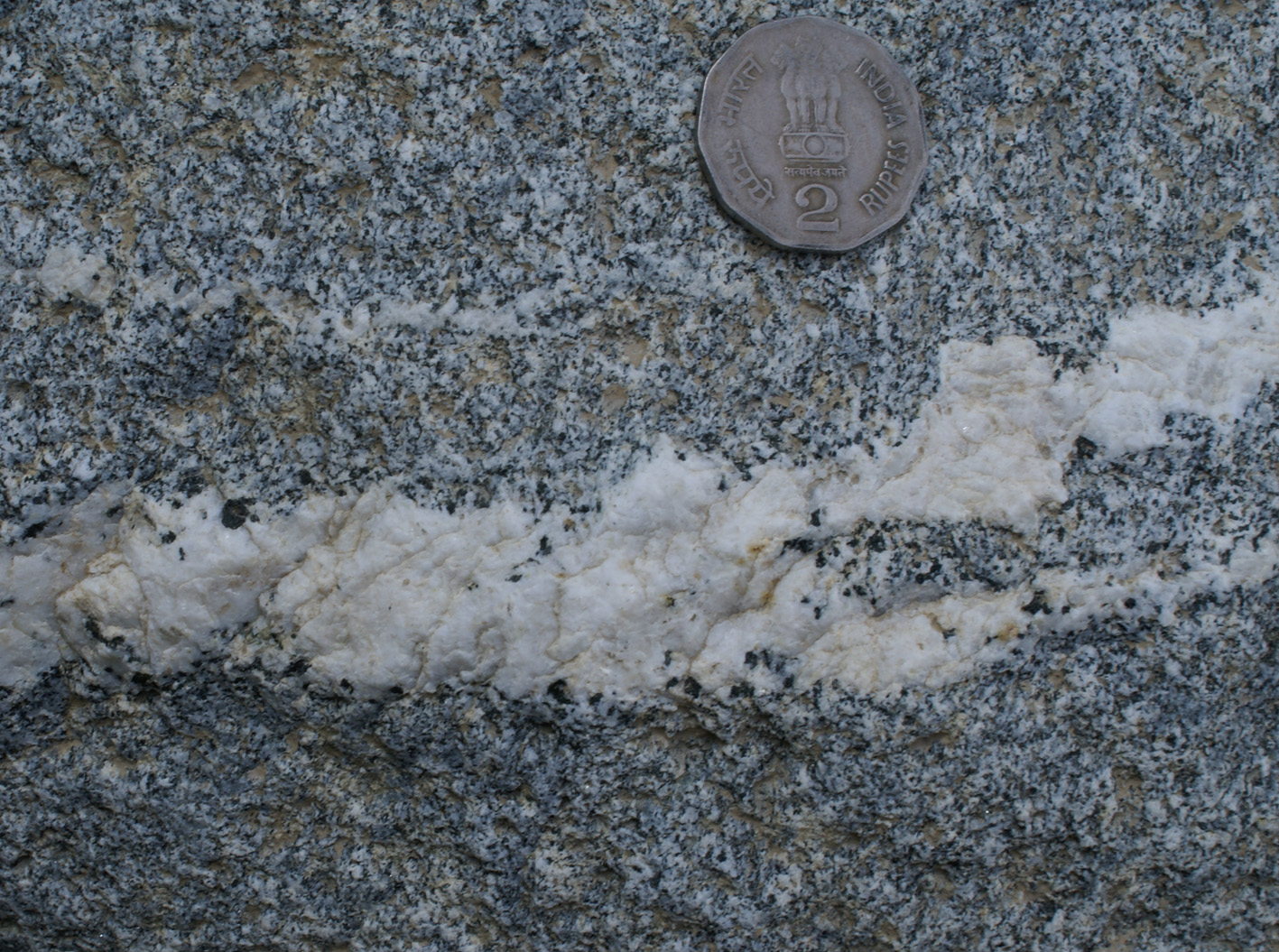
d) final product of melting 1003good
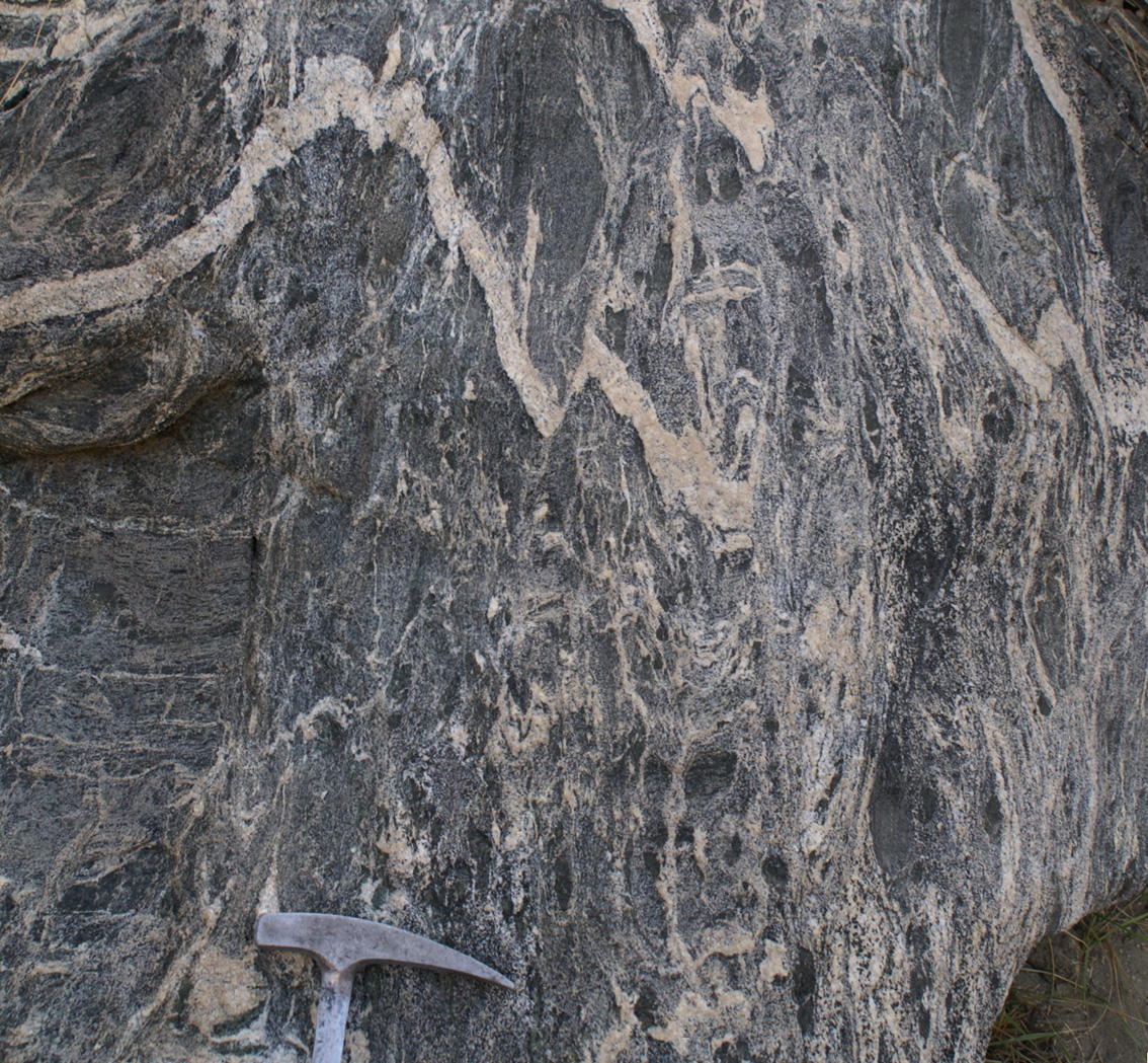
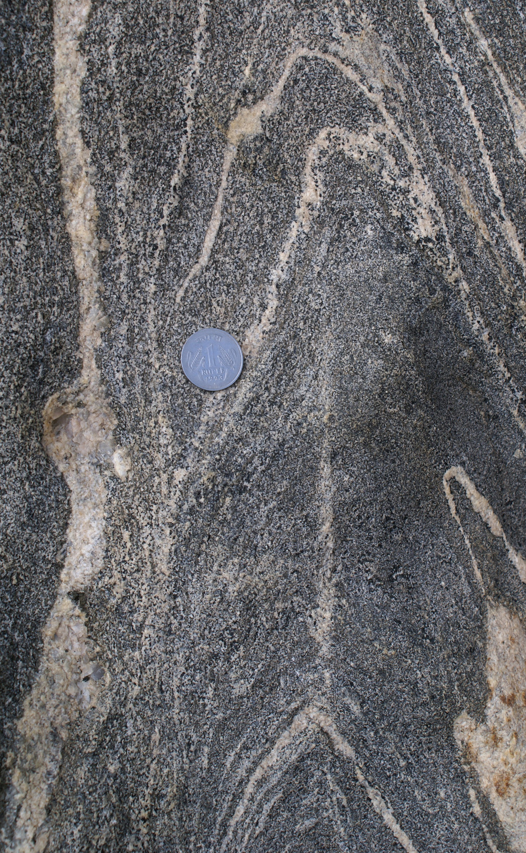
d) preference for antiform and destruction of synform, 4759 needs to be accompanied by a line drawing, 4779good, IMG_4776zoom, 4784good synform and melting pushing up, stitched_894, DSC00879good
a) pattern of melting, 4763, 4767, 4780, 4751 and detail in 4755 , 875, 933, 935, 915, 4770
b) break up of amphibolite to form diatexite stitch 925-926, show also 924, DSC00921good
c) folding and melting 885good, DSC00888zoom, 1216
d) final product of melting 1003good
d) preference for antiform and destruction of synform, 4759 needs to be accompanied by a line drawing, 4779good, IMG_4776zoom, 4784good synform and melting pushing up, stitched_894, DSC00879good
a) In situ
melting of biotite-amphibolite (all photographs down plunge of fold
axis, or perpendicular to axial plane)
|
|
|
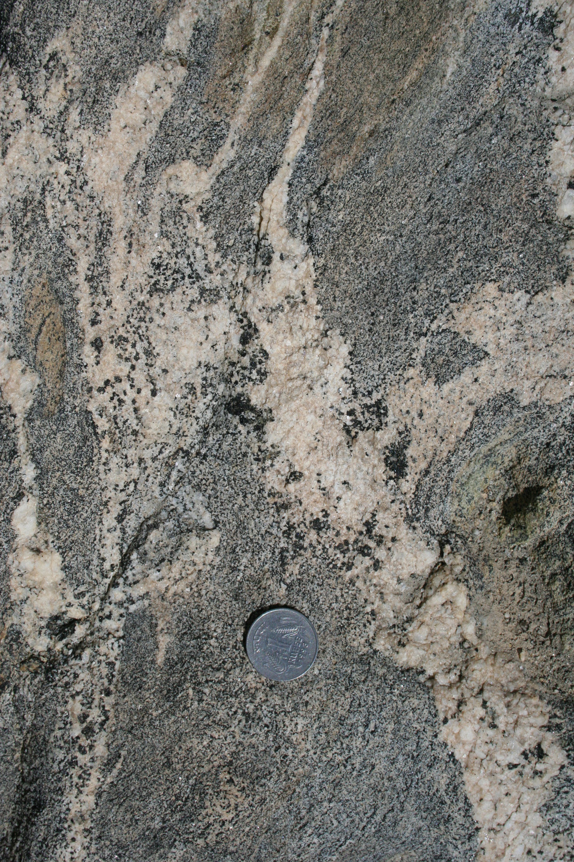
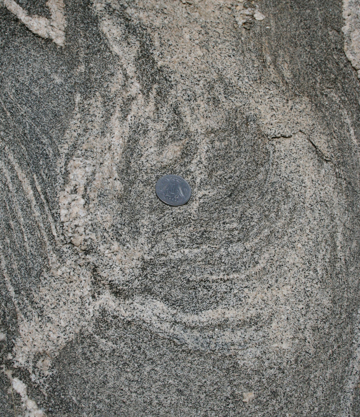
| Figure 2a-c. (4780, 4751, 4755) outcrop | Figure 2b. Folded amphibolite displaying leucosomes linked directly with the matrix as well as sharp leucocratic and folded dykes. (outcrop) |
|
|
|
| Figure 2c. Detail of upper right-hand-side of 2b, where leucosome displays a feathery pattern. |
|
|
|
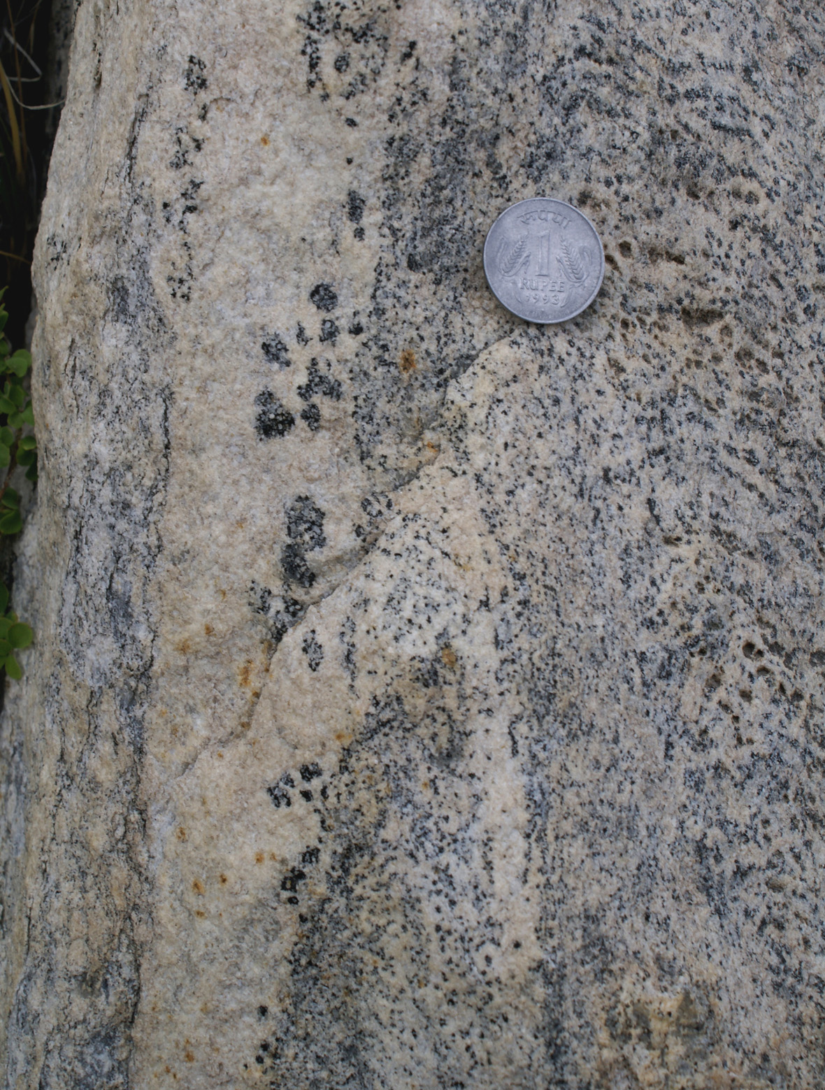
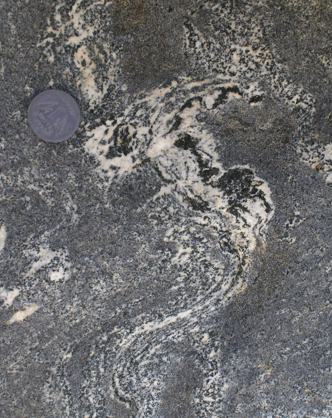
| Figure 3a-c. (933, 935, 915). In situ melting in amphibolite block forming leucosome surrouding hornblende phenocrysts. (block) | Figure 3b. Disaggregation of folded biotite-amphibolilte. Leucosome has hornblende phenocrysts showing preference to grow on isolated amphibolite block (upper right). (block) |
|
|
|
| Figure 3c. Partly preserved block of biotite-amphibolite surrounded by a narrow rim of leucosome and a hornblende diatexite. Note that the block itself has leucocratic patches, better developed on the right-hand side where its boundary with the surrounding is diffuse and internal leucosomes link up with the external groundmass. (block) |
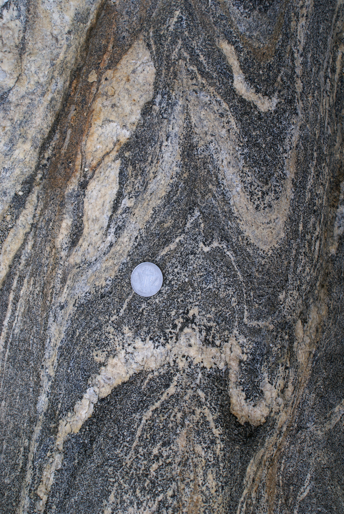
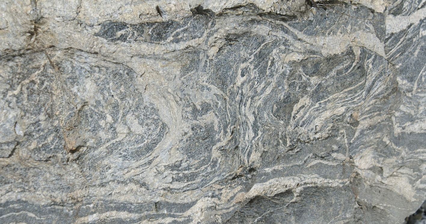
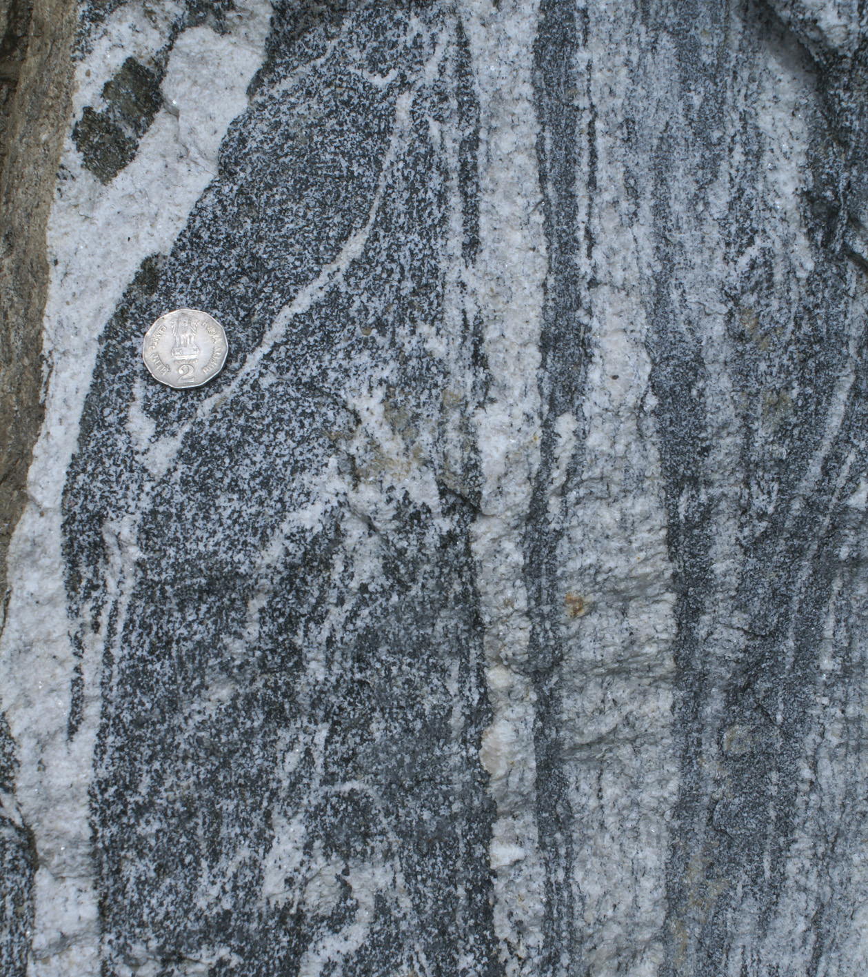
b) Amphibolite break-up: diatexite formation
 |
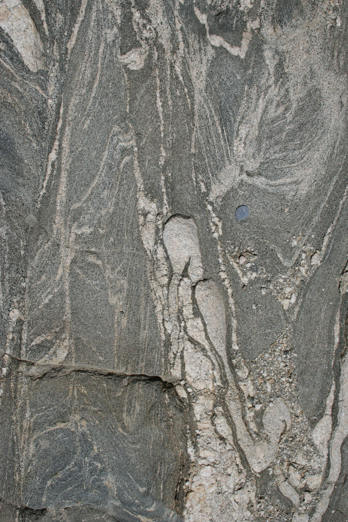
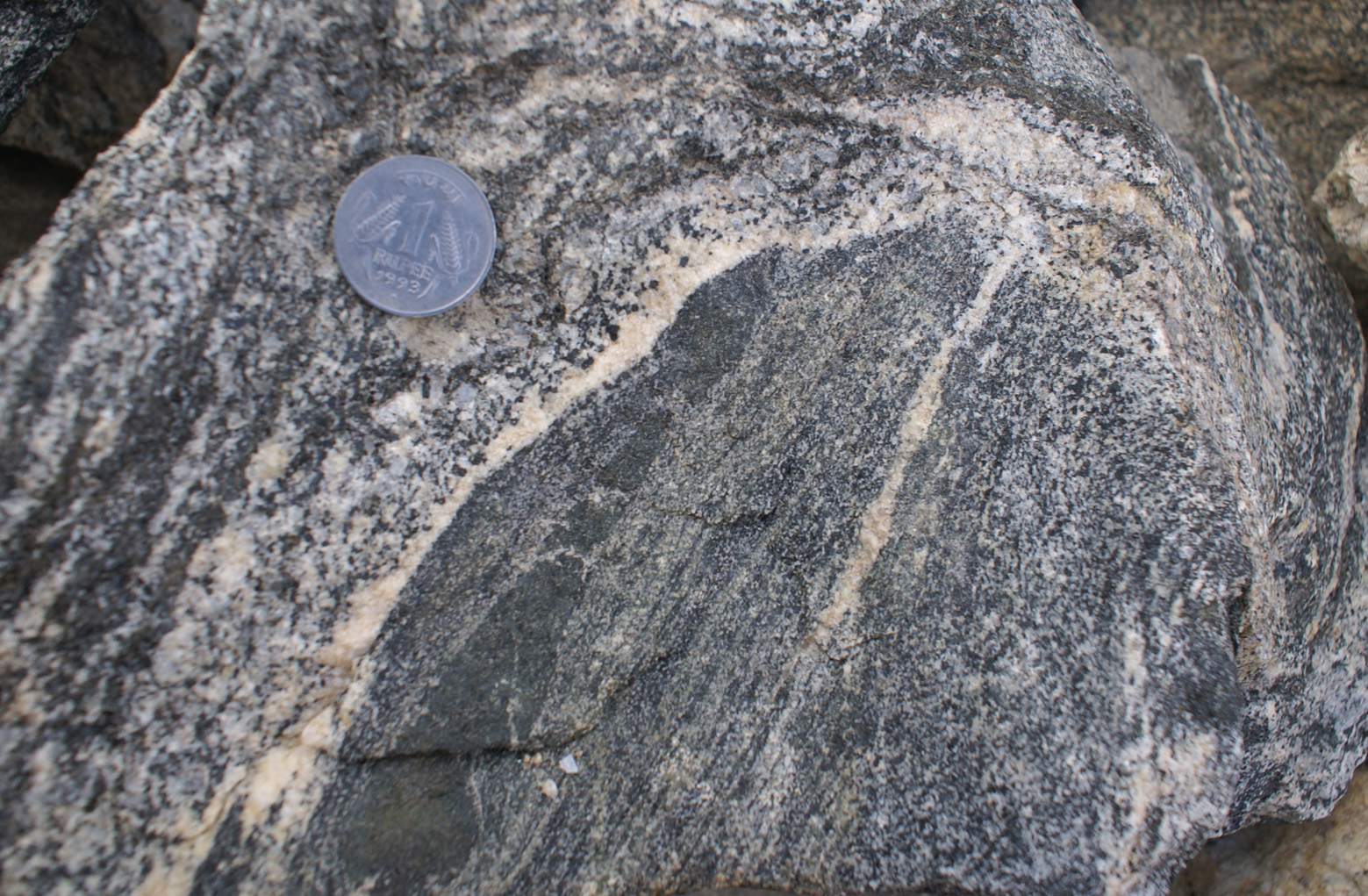
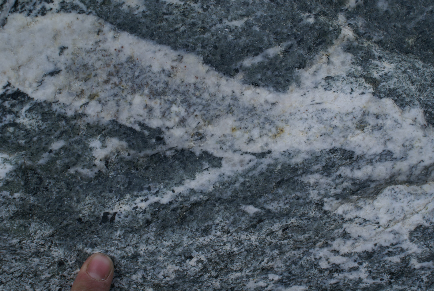
| Figure 4 (925-926): This block of amphibolite shows three stages of break up due to melting and melt migration. On the left-hand-side a metric block of amphibolite has an original foliation (horizontal in the photograph) and few leucosomes. The central part shows an increase in leucosome fraction and the partial disruption of the amphibolite. On the right-hand-side the original amphibolite is disrupted to form a diatexite. This evolution from left to right is traced by the leucocratic dyke in the upper part of the photograph, which evolves from relatively straight, on the left, to folded in the centre and disrupted on the right, and reappears in the far right again. |
|
|
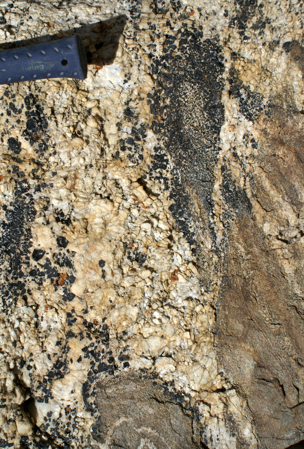
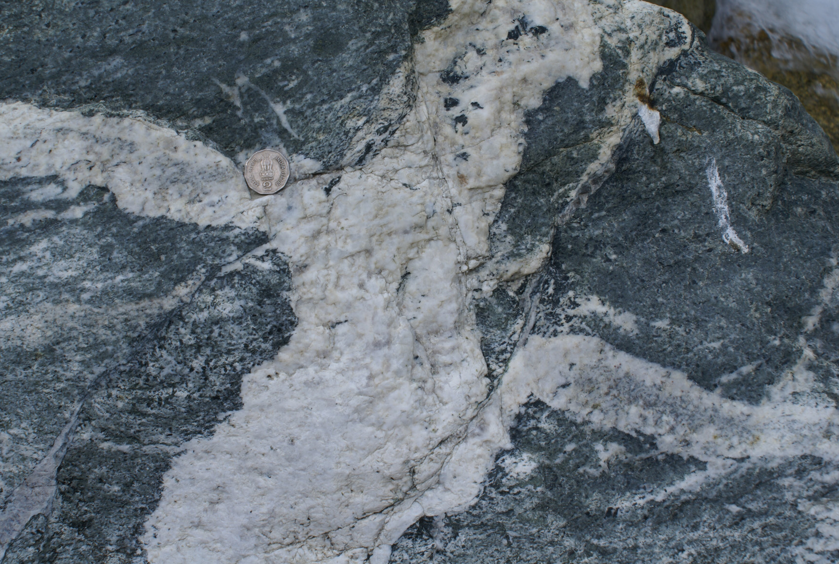
| Figure 5. Leuco or mesocratic irregular bands pervading the rock and isolating blocks of amphibolite. Note diffuse boundaries everywhere. Interpretation: melt channelway disaggregating the source rock and mobilizing magma: (block surface slightly curved, photo at a high angle to the leucocratic bands but due to variation in strike and block curvature there is some cut effect). |
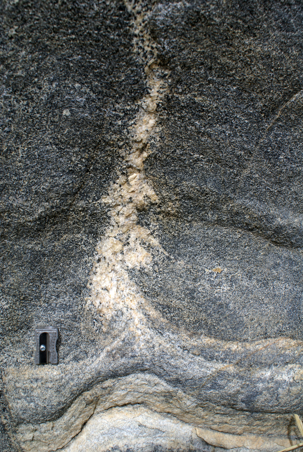
c) Folding and axial planar leucosomes
|
|
|
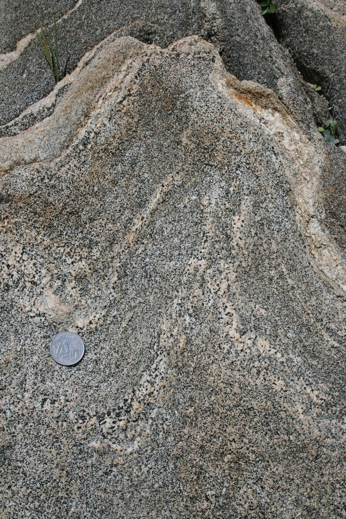
| Figure 6. (885good). In situ melting in amphibolite during folding (outcrop) | Figure 6b (888zoom). Escape channelway of melt along the axial plane inferred from the narrow diffuse leucosome through the hinge zone. (outcrop) |
 |
|
| Figure 6c (1216). Incipient folding triggering axial planar leucosome accumulation. |
d) Final product of melting
|
|
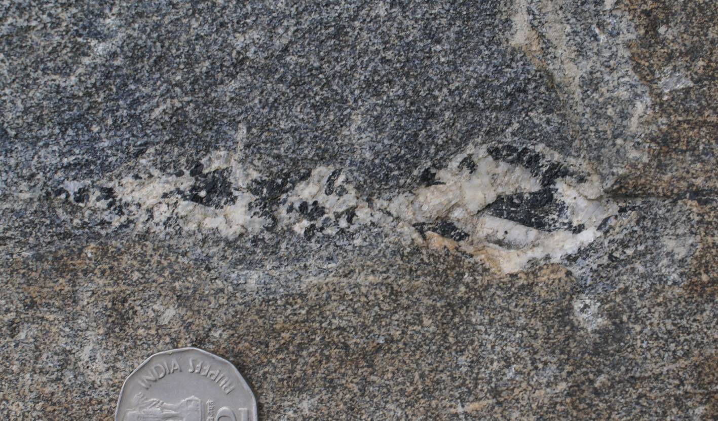
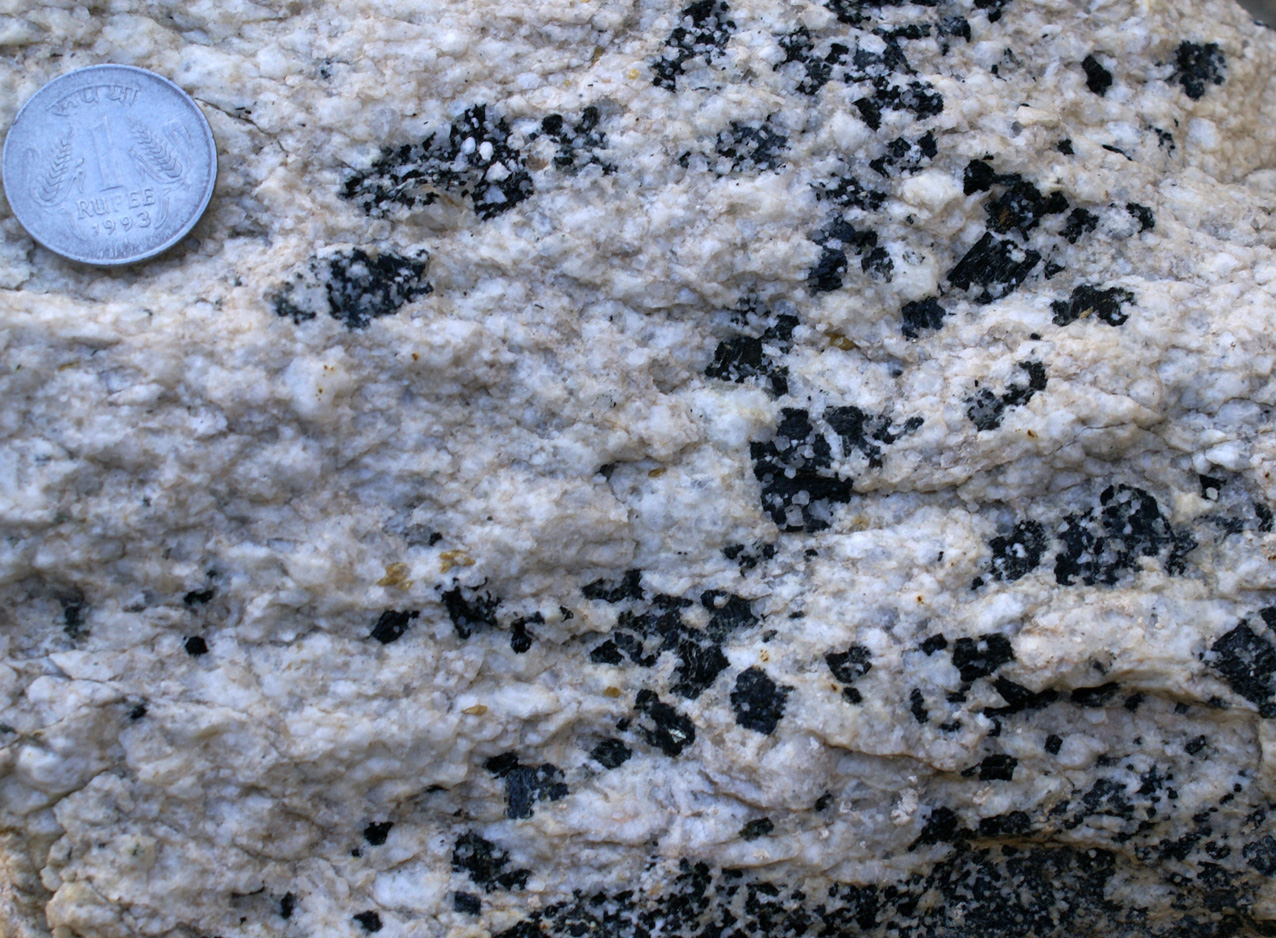
| Figure 7. Typical melting product of amphibolites:: leucocratic rock with poikilitic porphyroblasts of hornblende (inclusions generally quartz). This fabric was used elsewhere in the region to recognize partial melting in deformed rocks. |
e) Examples elsewhere along the Pangong Range
Shyok - Lomo Yogma, 20 km SE of Agham
|
|
|
| Figure 8a,b,c. Lomo Yogma valley. Leucogranite dykes with hornblende phenocrysts (upper, central part). Dykes have irregular boundaries with intermediate biotite amphibolite (see zoon in b). | Figure 8b. Detail of lower right-hand-side of b, showing continuity of leucogranite dykes down to groundmass scale, and the gorwht of a porphyroblast in the matrix, immediately above the finger. |
|
|
|
| Figure 8c. Leucograntie with irregular boundaries against intermediate biotite-amphibolite, and containing hornblende porphyroblasts preferentially close to the margin. |
Shyok - Darbuk Gorge
|
|
|
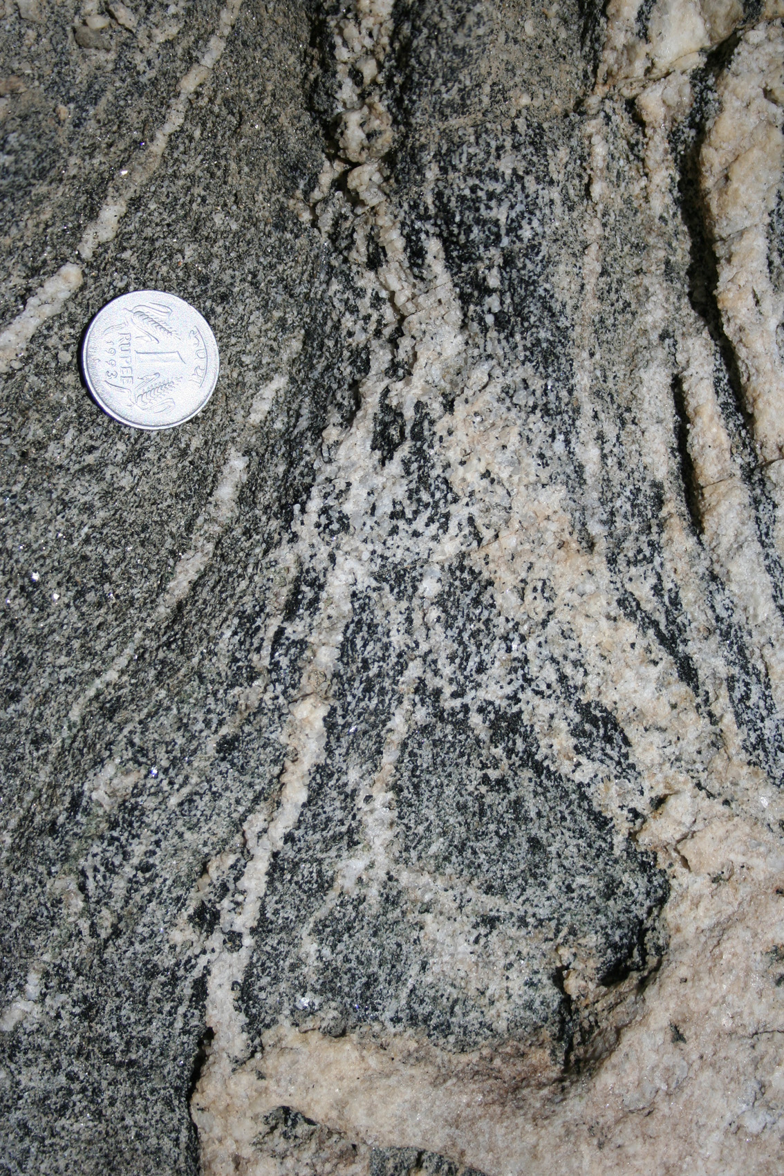
| Figure 9. Diatexite in Bt-Hbl-bearing gneiss, presumably related to water-present melting with production of peritectic hornblende (Fig. 9b). Length is 3m. | Figure 9b. Same outcrop as 9a. In situ melting (center of photograph) giving rise to a leucogranite and porphyroblasts of euhedral, >2cm Hbl (upper left-hand-side). |
|
|
|
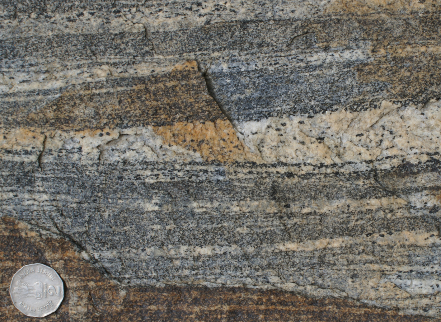
| Figure 9c.Same outrcrop as b,c, showing leucogranitic material surrounding poikiloblatic Hbl. | Figure 9d. Typical banding comprised of mafic Bt-Hbl gneisses and leucocratic bands with porphyroblasts of Hbl. |
|
|
|
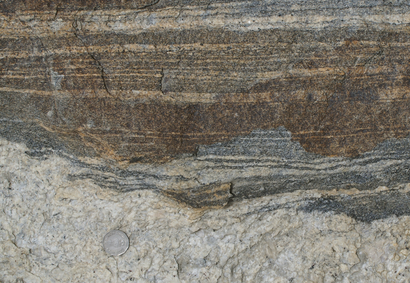
| Figure 9e. Continuous merging between layer parallel leucocratic bands and cross-cutting leucogranite with rare mafic grains. |