Overprinting Anatectic Events, High Himalaya Crystalline, Sikkim
|
| This site describes two outcrops in the High Himalayas Crystallines in Sikkim, where there is possible evidence for two anatectic events. |
Outcrop 13. After Bonsoi Bridge, N27 42'14.0" E88 33'44.9".
This outcrop is an orthogneiss migmatitic sequence, with a 2m wide migmatitic amphibolite layer, a 1m wide intrusive granite, all parallel to a dominant migmatitic foliation trending 340/60E (Fig. 1 b to g). This is overprinted by a 2m wide deformation zone, trending N70E/sv (Fig. 1a). This has a pervasive foliation, tight folds with 10-15cm thick mylonite, intrusive leucogranite dykes and in situ leucosomes seggregated to form leucosomes parallel to the axial plane of the folds. This is a possible example of two melting events. We have collected a sample SK13-6 of the late granite intrusive, that dates the second melting event and the mylonite that cross-cuts earlier, dominant foliation. This outrcop also has evidence of amphibolites undergoing partial melting during the first melting event, with Grt and possible Px (now retrogressed) associated with leucosome. A possible example of the prediction of high T melting during the Himalayan Orogeny (Thompson and England 1986).From Rubatto et al. 2013 (CMP p. 357) for sample CLN6D a metapelite from the same outrcop (? N27 42.247 E88 33.727): "Monazites in metapelite CLN6D have two domains that are distinct in zoning, chemistry and age. Unzoned to weak oscillatory domains (cores) yielded dates between 29 and 27 Ma that define a broad peak at 27.8 +/- 0.3 Ma (MSWD 2.3). In numerous grains, the cores are cross cut by rims with patchy, irregular zoning that yield younger dates between 25.3 and 22.5 Ma. Chemically, the crosscutting rims are distinctly higher in HREE and Y contents and have a slightly stronger Eu anomaly (Fig. 5). Notably, a few monazite cores contain either K-feldspar or K-feldspar+quartz inclusions."
Rubatto et al. (2013) sugggest that these two age groups reflect two melting events, one during prograde metamorphism accompanied by the growth of Grt forming Mnz with low values of HREE. This early event was followed by a second melting event during decompression leading to monazites with higher HREE indicative of retrogression of Grt, liberating HREE. If this is the case, regional peak metamorphism was associated with the NNW-trending foliation and the later N70E overprint formed during decompression a few million years later.
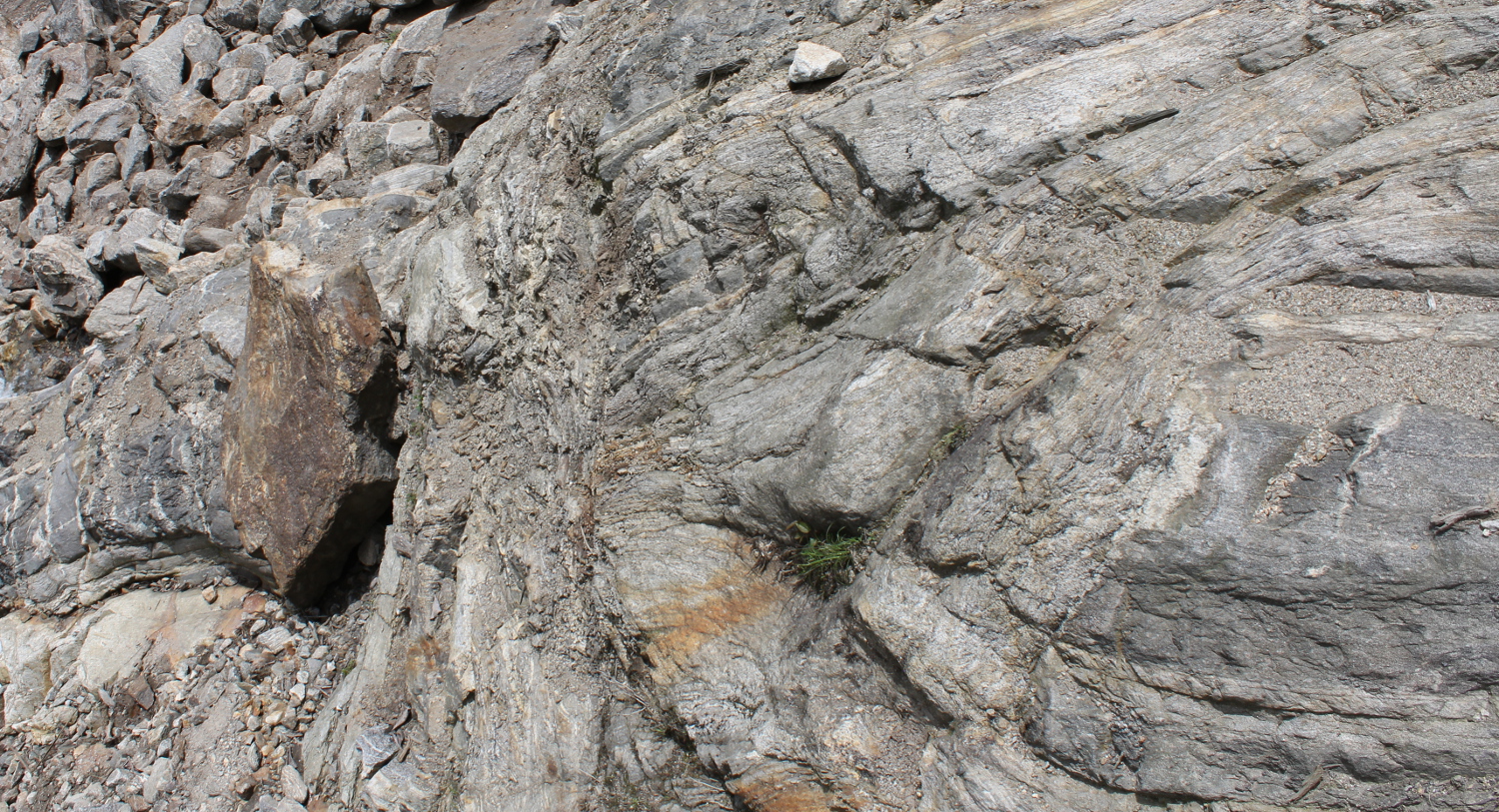
Fig. 1a) The main outrcop section showing a boudinaged amphibolite layer in the foreground (lower right) surrounded by gneiss, and at the far end (left). Details of amphibolite shown in Fig. 1c, d. There is a 2m wide zone of shearing trending at a high ange to the dominant foliation associated also with granite intrusion, trending N70E/sv. |
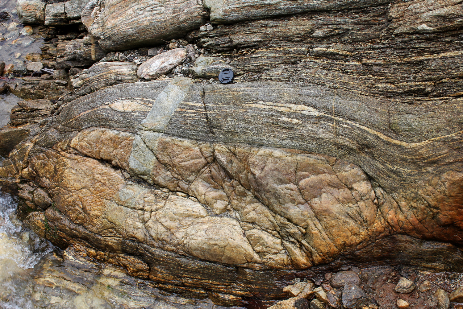
Fig. 1b) Boudin of leucogranite parallel to S1, cut by a late dyke. |
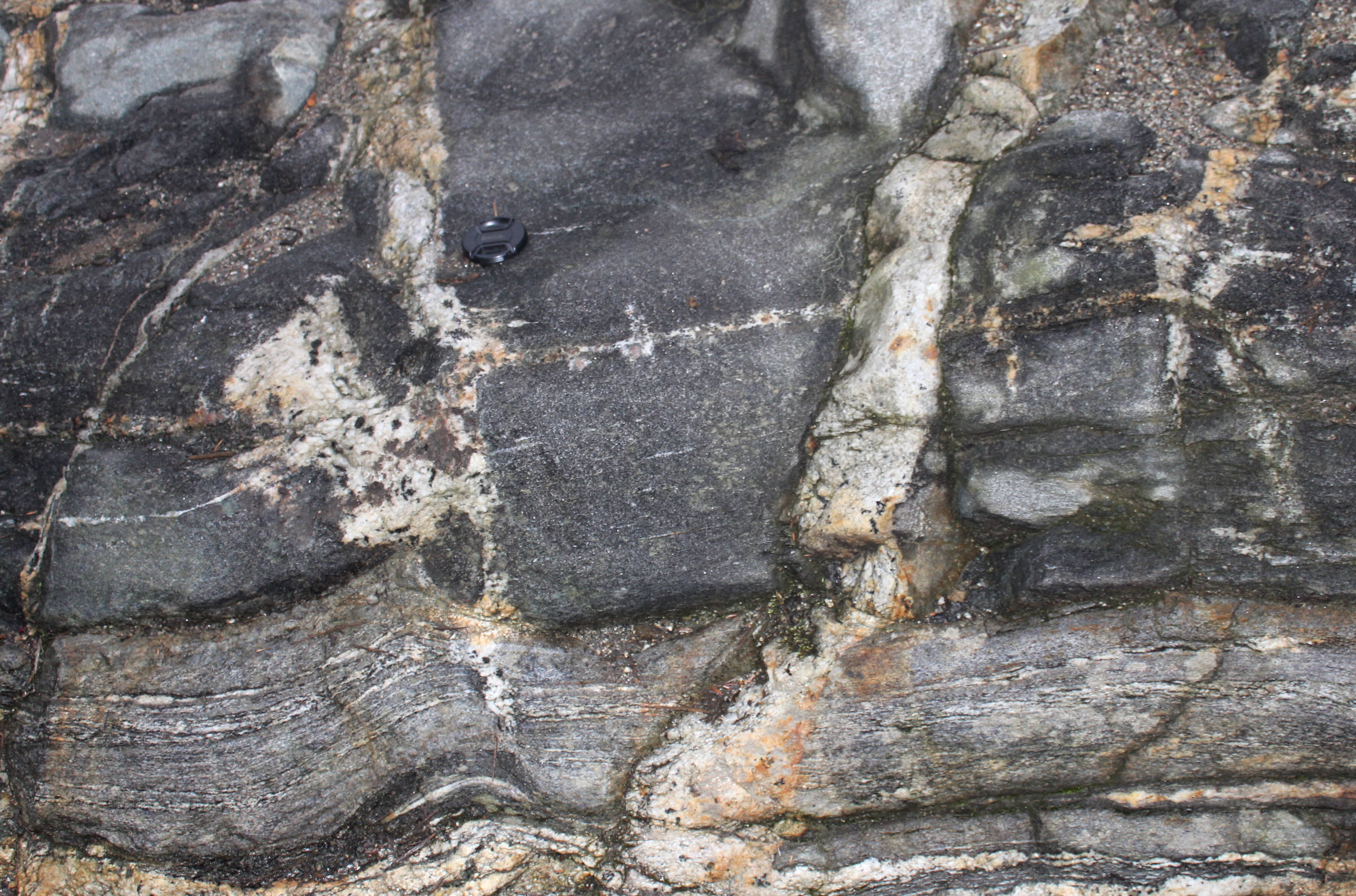
Fig. 1c) Leucosome with large garnet in amphibolite, cut across by a pegmatite dyke. |
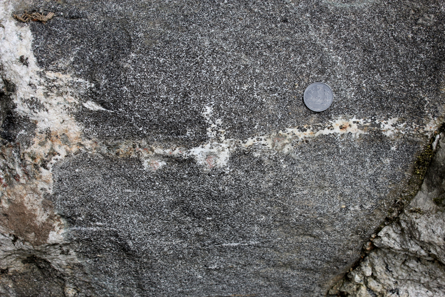
Fig. 1d) Detail of Fig. 1c, in situ melting of amphibolite forming a Grn-Hbl-Bt-(Ms-Chl??) leucosome with irregular margins marging into surrounding amphibolite. |
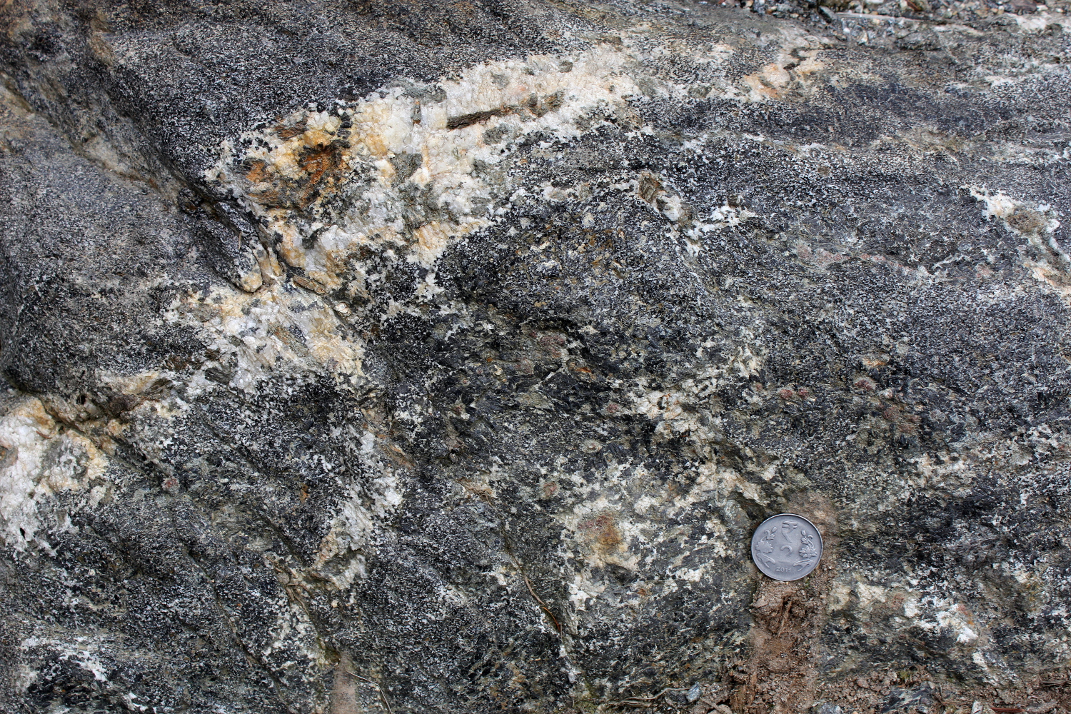
Fig. 1e) Leucosome in amphibolite with large garnet with a greenish rim of Pl and Bt(?) and a leucosome with large Hbl interpreted to be peritectic. |
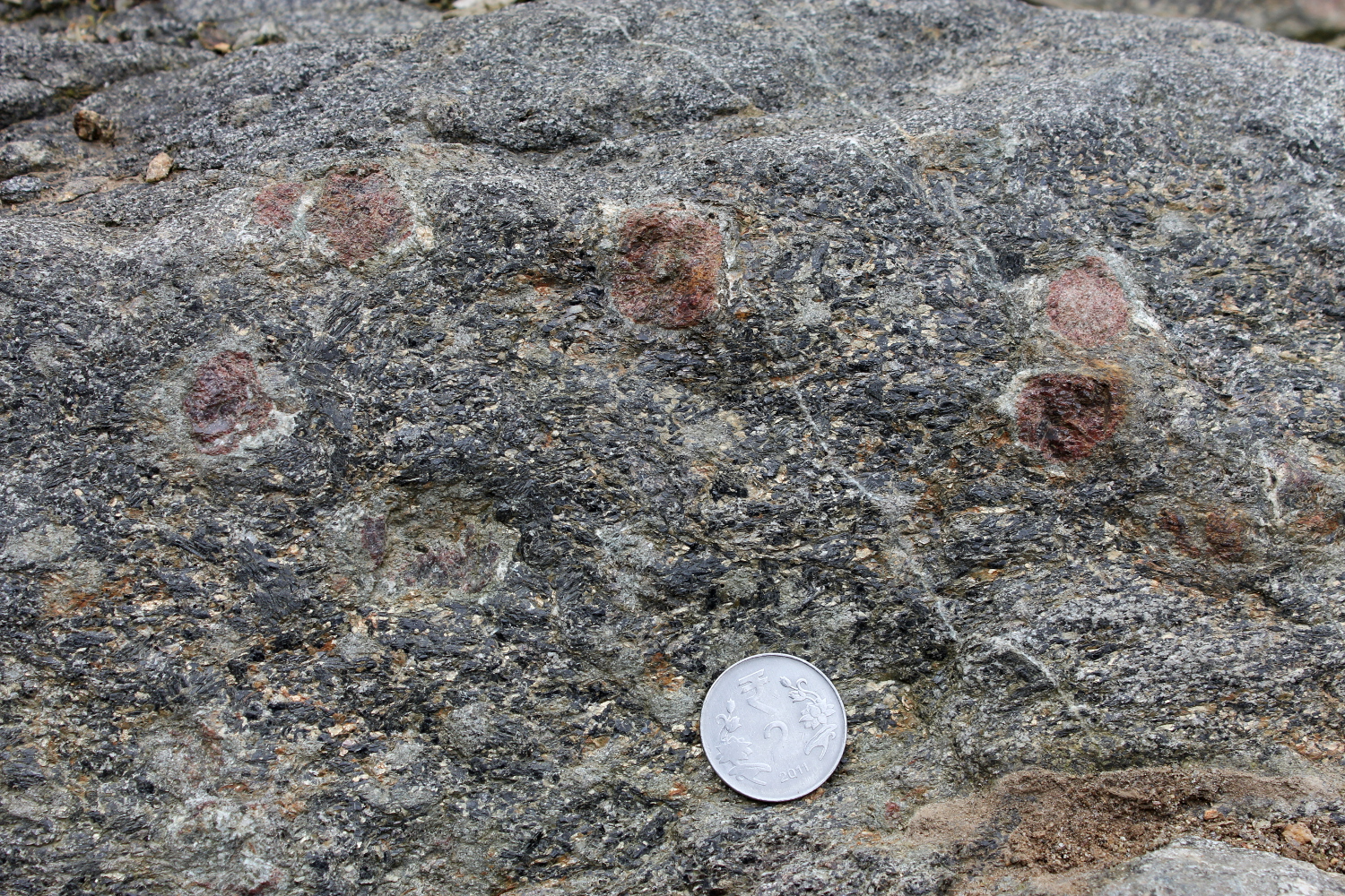
Fig. 1f) Garnet, 2cm diameter, with strain shadows of very fine Pl-Bt asymmetric tails in amphibolite. Interpretation: residual rock comprising peritectic garnet and possibly peritectic hornblende. |
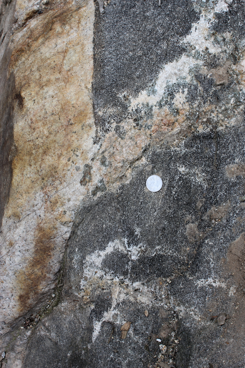
Fig. 1f) In situ leucosome merging with late intruded leucogranite with sharp boundaries against amphibolite. Leucosome has Hbl (black), Bt and square and rectangular light green pseudomorphs after Hbl? Or Opx? Possibly now tremolite-actionolite. |
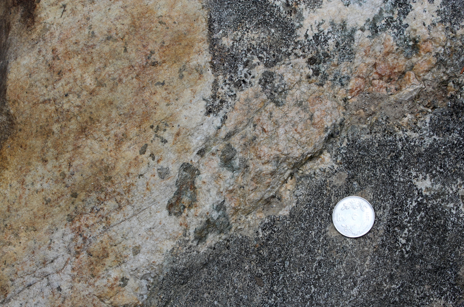
Fig. 1g) In situ leucosome merging with late intruded leucogranite with sharp boundaries against amphibolite. Leucosome has Hbl (black), Bt and square and rectangular light green pseudomorphs after Hbl? Or Opx? Possibly now tremolite-actionolite. |
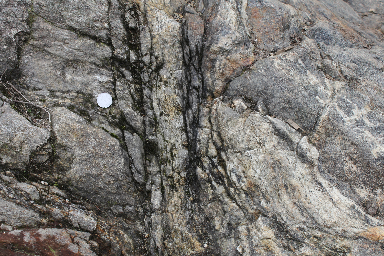
Fig. 2a) Shear zone trending N70E/sv, vertical exposure seen parallel to lineation, perpendicular to foliation, south side down. |
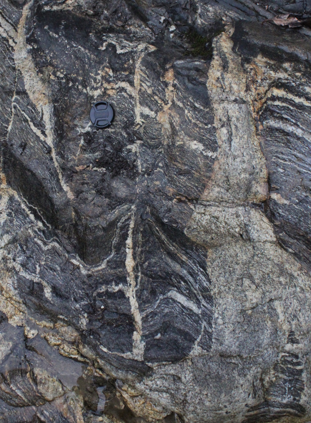
Fig. 2b) Leucogranite dykes intruding N70E/sv planes, parallel to mylonite zone in Fig. 2a, cross-cutting gneissic foliation and folding it. Notice in situ leucosome in hinge zone of small fold. Dyke on left cross cuts leucosomes. Detail in Fig. 2c. |
Folded cross-bedding in quartzite. Notice truncation of layers on the left by the darker layer. Pandukeshwar Fm on the way to Malari, HHC. |
Outcrop 34.
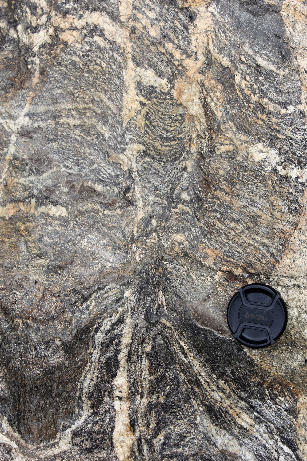
Migmatite gneisses with an early foliation with leucosomes, folded and sheared during anatexis. The interesting aspect of this outcrop is the close relationship between melt segregation and folds and shear zones (Fig. 3). Melt segregates into shear zones or axial plane of folds. Folds with such leucosomes may grade into shear zones, or leucosomes and these structures may disappear along strike. This outrcops also show a that there are possibly two anatectic events (Fig. 4). N65-70E/sv Lst=000/55, and folds, N27°38'19.8" E88°42'42.7" at a waterfall on the side of the Lachun road.
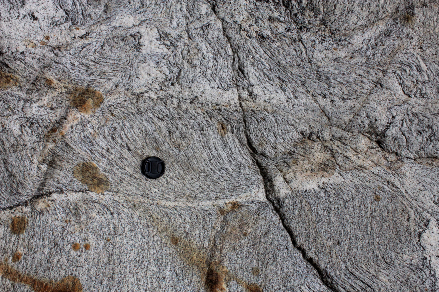
Fig. 3a) Incipient sinistral shear zone (in 2D) filled with a leucosome being fed directly from the surroundings. |
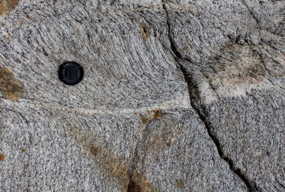 Fig. 3b) Detail of previous. |
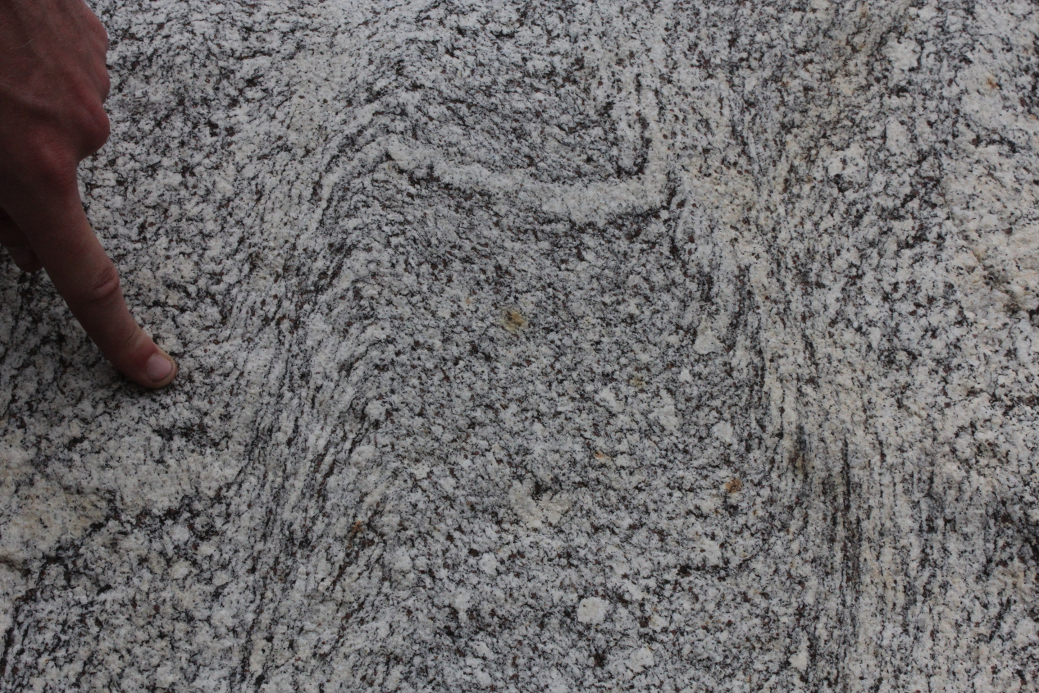 Fig. 3c) Two shear zones bounding a lithon of weakly deformed gneiss |
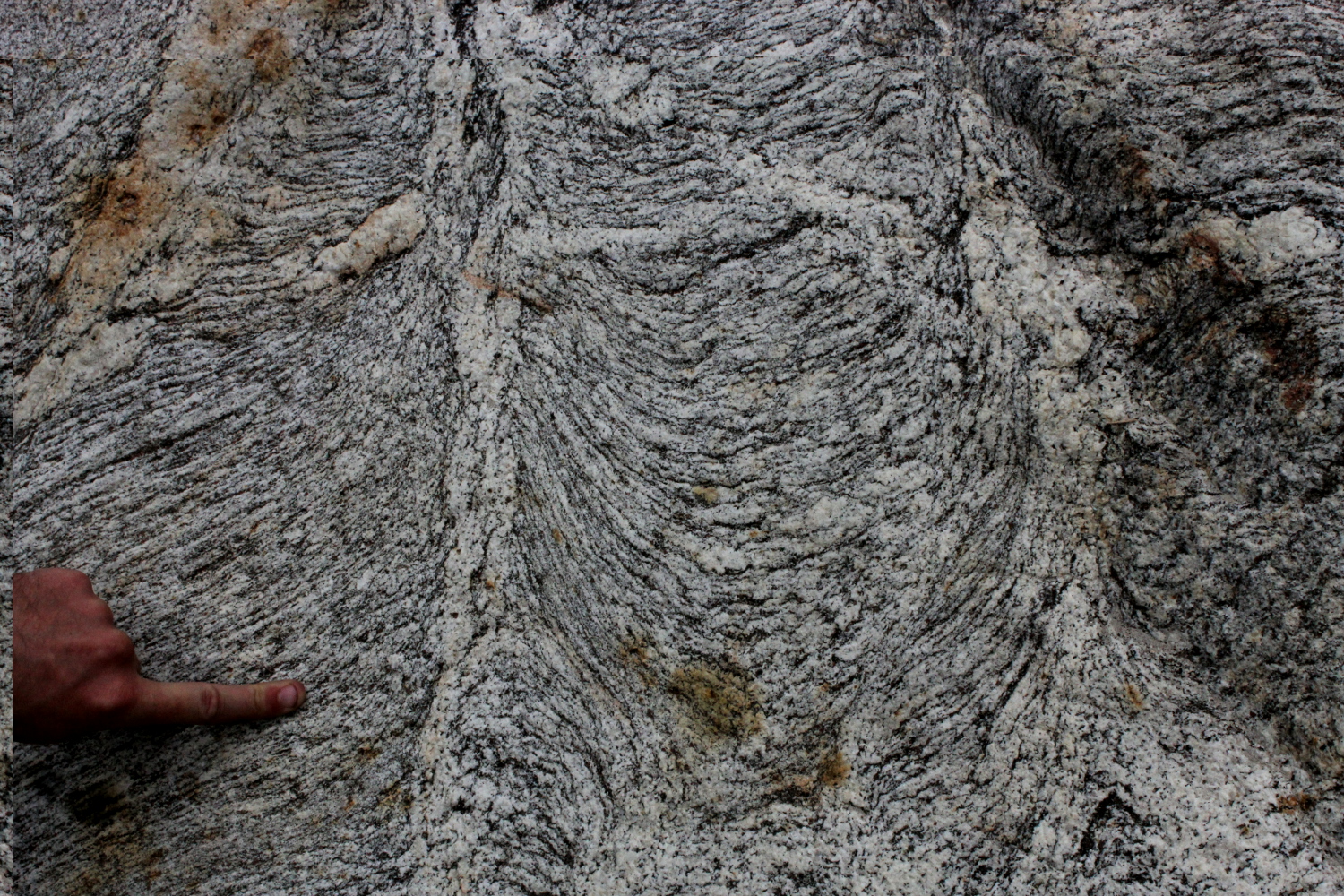 Fig. 3d) Two axial planar leucosomes in two folds. On the left, leucosome is in cuspate unidirectional fold hinge. Note it develops from the hinge of an antiform. On the right, the leucosome develops from the top of an antiform as well but further up this becomes a shear zone. |
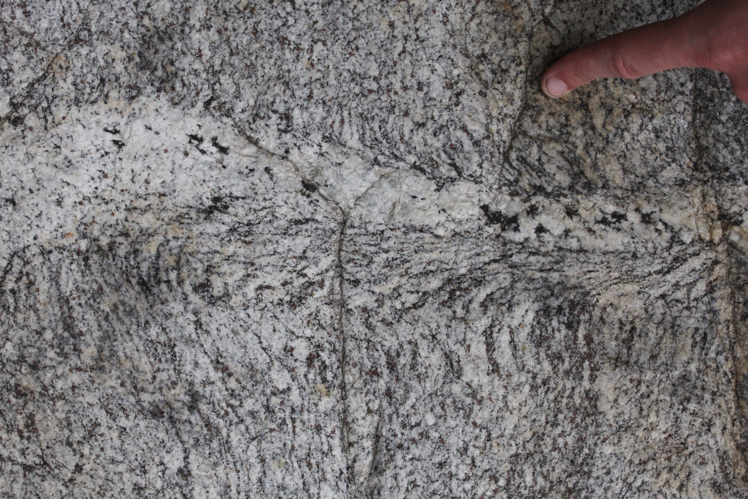 Fig. 3e) In situ leucosome in axial plane of a cuspate fold |
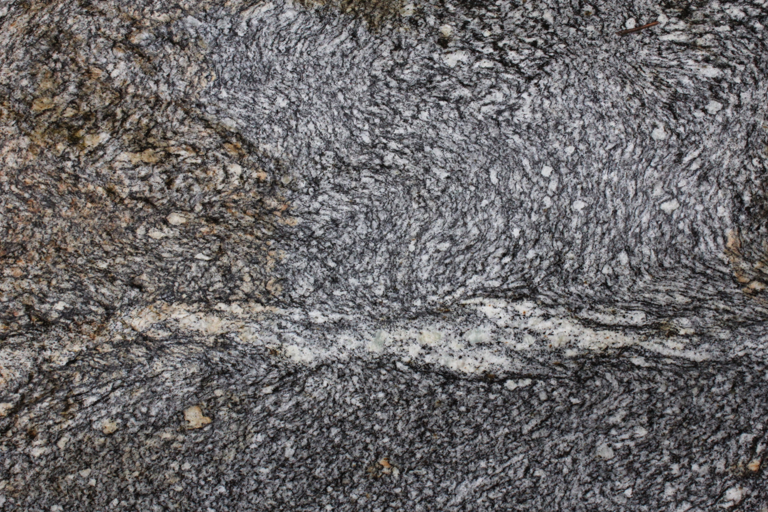 Fig. 3f) In situ leucosome/melanosome in axial plane of a cuspate fold. |
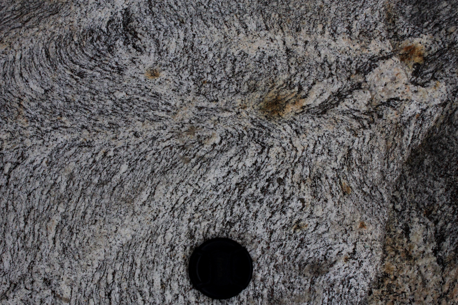 Fig. 3g) Three in situ leucosomes converging towards a fold hinge. |
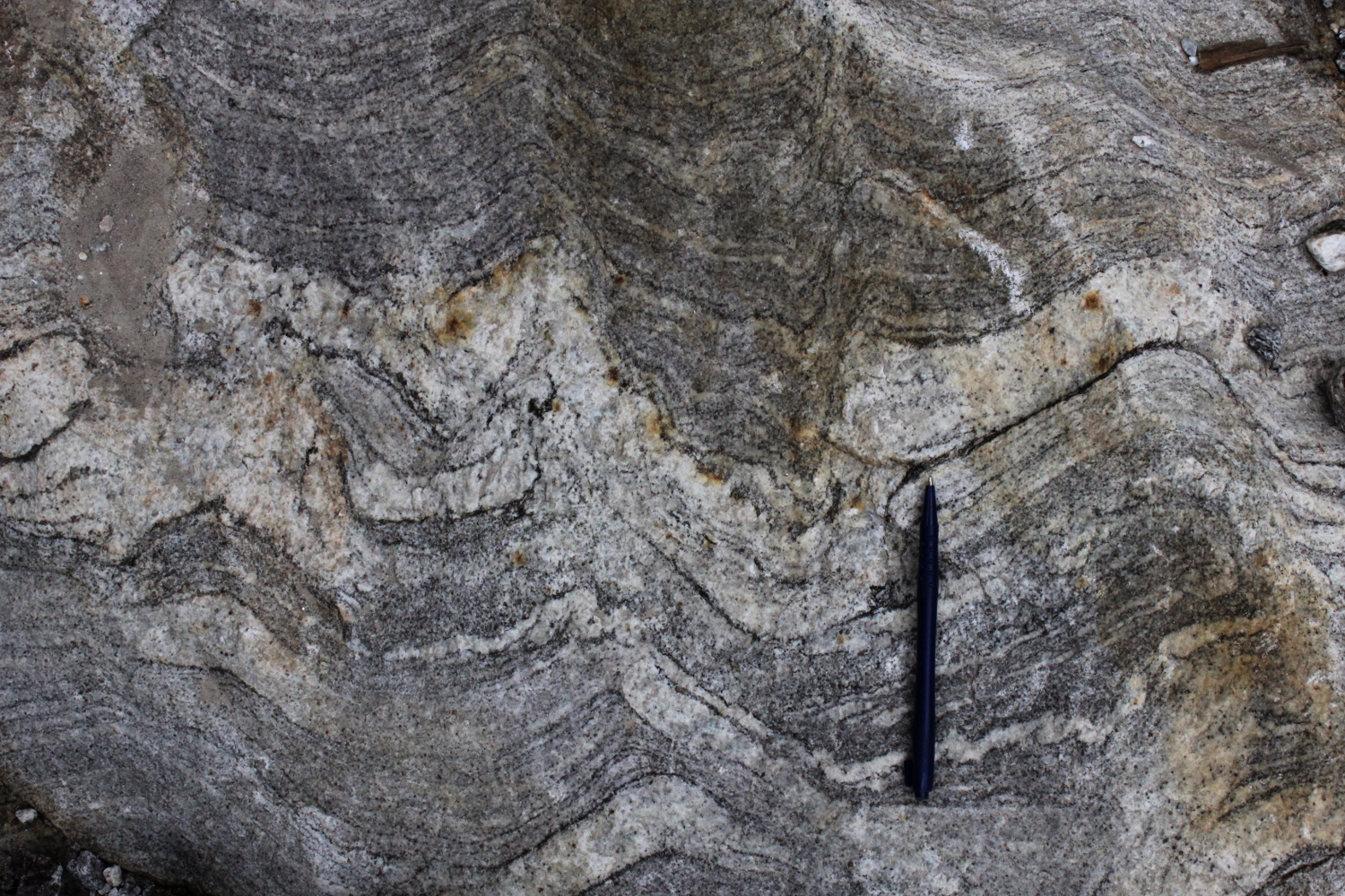 Fig. 4a) Migmatitic orthogneiss with stromatic leucosomes and melanosomes. This layering is blurred in the hinges of folds by a leucosome with fuzzy margins. Notice that the fold on the right-hand-side is not disturbed by leucosomes, but there are incipient leucosomes just starting to form above the leucocratic layer. |
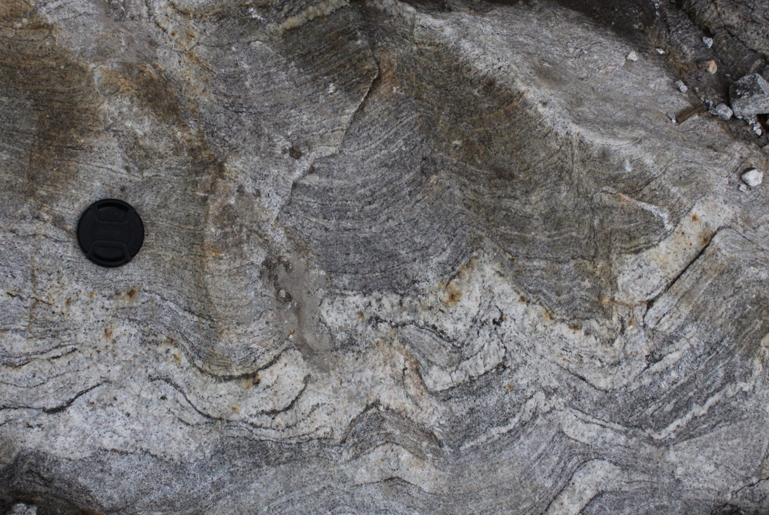 Fig. 4b) Banded leucogneiss with layering in fold hinges disturbed, truncated by new leucosomes. |
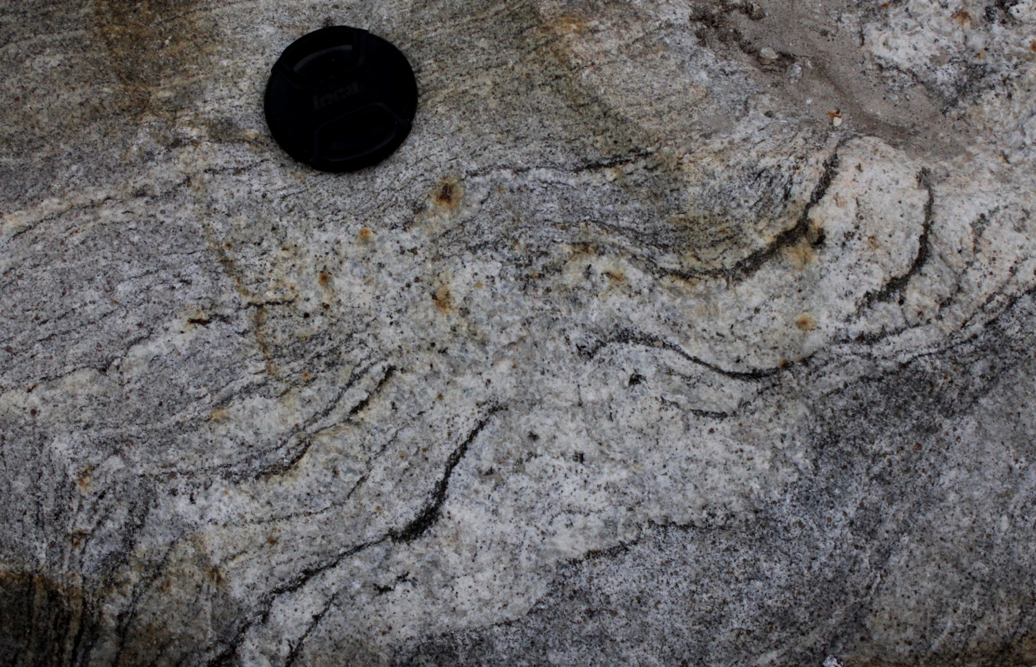 Fig. 4c) Detail of Fig. 4b showing the disrupted fold hinge. |
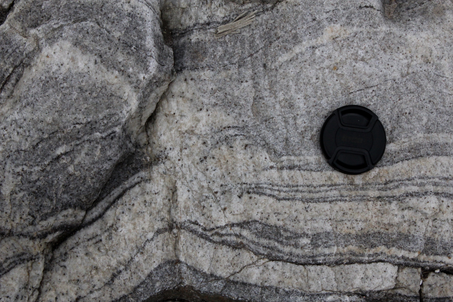 Fig. 4d) Banded leucogneiss with layering in fold hinges disturbed, truncated by new leucosomes. |
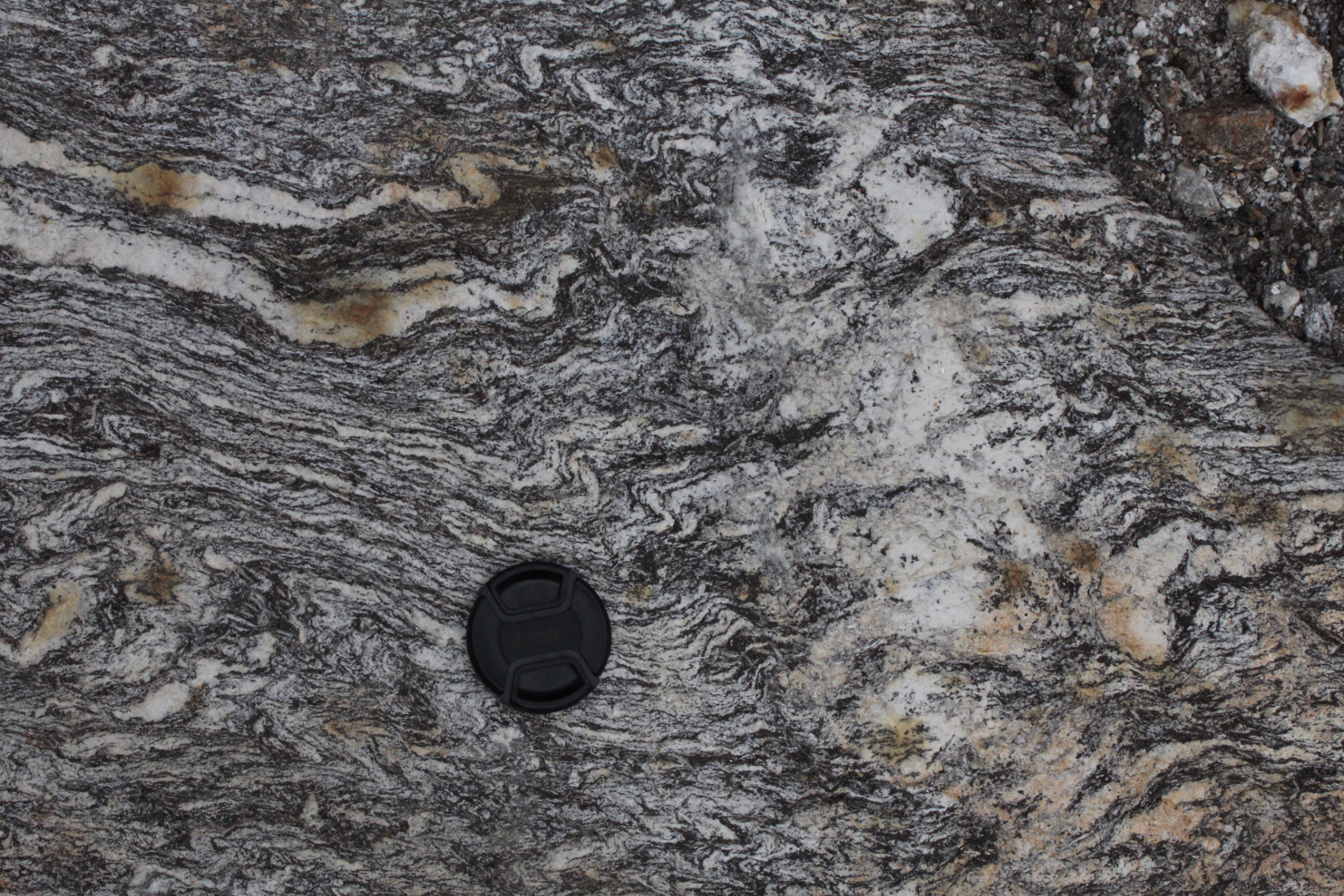 Fig. 4e) Migmatite overprinted by a new diffuse leucosome with Sil+Bt+Ms in contrast to Gnt in old leucosome. . |
Outcrop 8.
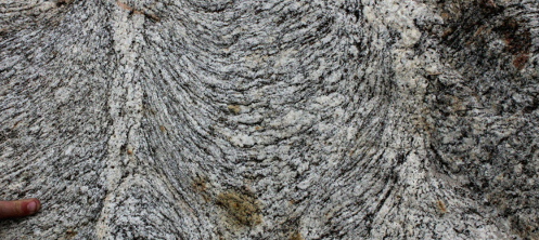
Beautiful strongly lineated migmatitic Ms-Bt granite, with leucosome rods. L=350/28, no good S-C fabric but sense is dextral thrust to S. N27°30'51.5" E88°34'39.4" road from Mangan towards Lachun.
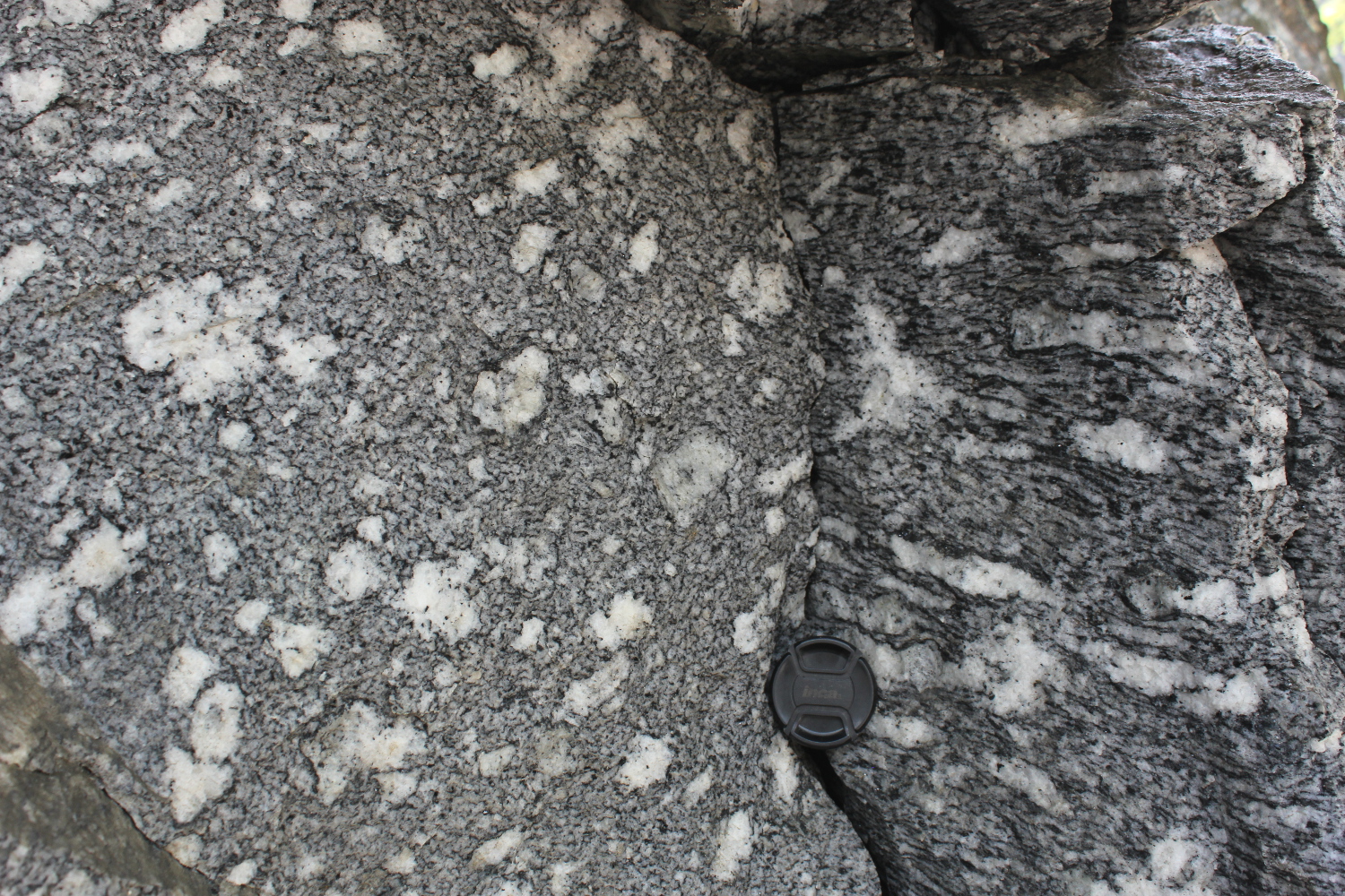
Fig. 5a) Bt-Ms orthogneiss with leucosome patches strongly elongated evidenced in one plane, with equidimensional cross-sections in the other plane. Notice melanosome. |
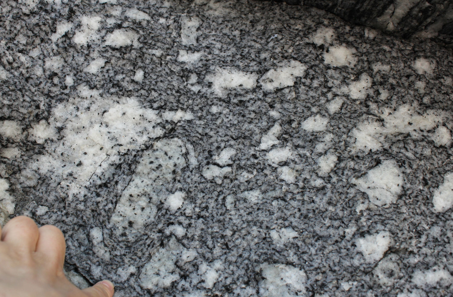 Fig. 5b) Leucosomes seen up plunge of lineation notice one with an apparent rotation marked by circumcentric foliations. |
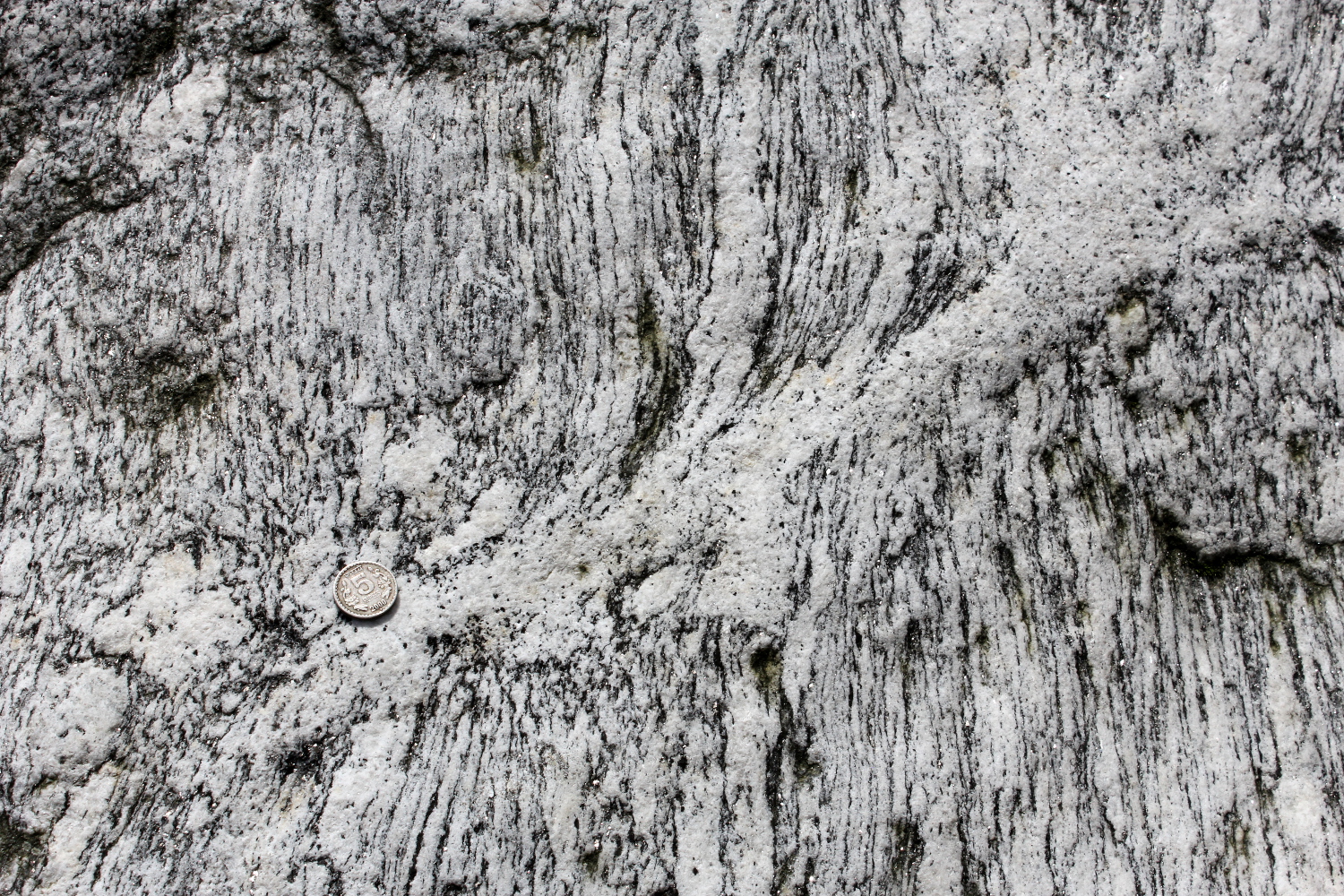
Fig. 5c) Same lineated migmatite with shear zone draining leucosomes. Plane parallel to L and perpendicular to S (xz plane). |
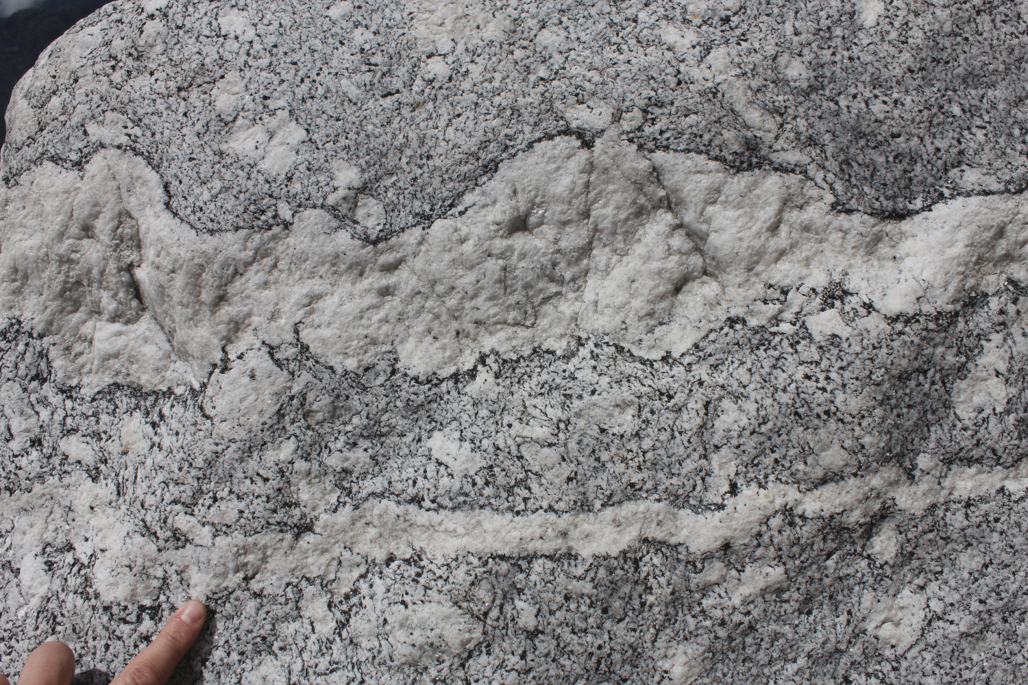 Fig. 5d) Irregular dyke with a narrow melanosome, looking down on lineation. |
Pt 23.
Migmatite Crd-Gnt-Sil-Bt paragneiss, photos looking updip of lineation and showing the single point accumulation of leucosomes drainage: a collapse feature. N27°54'16.4" E88°31'05.9" road N of Lachun, in Chopta valley past Thangu, 4700m altitude.
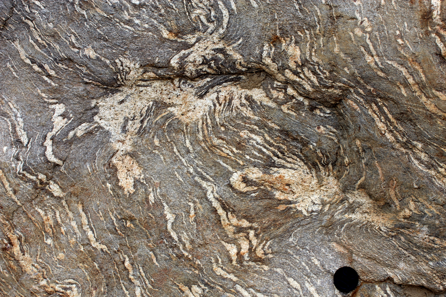
Fig. 6a) Accumulation and extraction of leucosome forming a collapse feature. Looking down plunge of the lineation. |
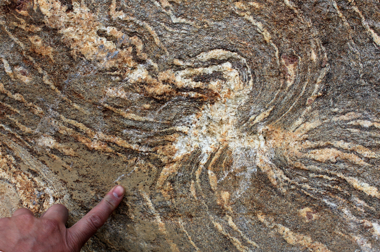 Fig. 6b) Accumulation and extraction of leucosome forming a collapse feature. Looking down plunge of the lineation. |
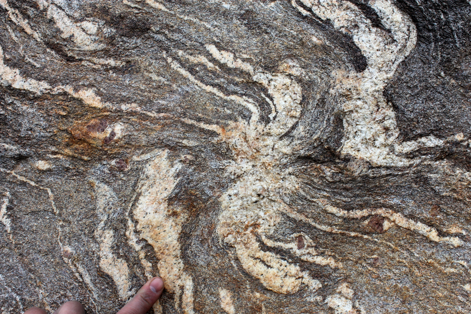
Fig. 6c) Accumulation and extraction of leucosome forming a collapse feature. Looking down plunge of the lineation. |